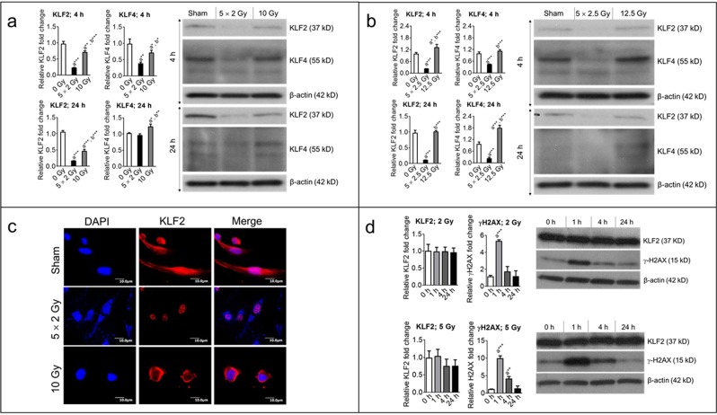
Figure 1.
Fractionated, compared to single exposure, radiation profoundly suppresses levels of KLF2 and KLF4. Representative (3–5 independent experiments) Western blot analysis and quantification of KLF2 (n = 5) and KLF4 (n = 3) levels in whole-cell lysates from nonirradiated (sham) and irradiated HUVECs 4 h and 24 h after exposure to (a) either five fractions of 2 Gy (5 × 2 Gy) or single exposure to 10 Gy and (b) either five fractions of 2.5 Gy (5 × 2.5 Gy) or single exposure to 12.5 Gy. Fractions delivered at 24-h intervals. β-actin served as a loading control. c Representative photomicrograph (20× magnification) showing immunofluorescence of KLF2 (red) in nonirradiated (sham) and irradiated HUVECs 4 h after exposure to either five fractions of 2 Gy or single exposure to 10 Gy (n = 3). Nuclei were stained with DAPI (blue). d Representative Western blot analysis and quantification of KLF2 (n = 3) and γ-H2AX (n = 3) phosphorylation levels in nonirradiated (0 h) and irradiated HUVECs at indicated time intervals after single exposure to 2 Gy and 5 Gy. β-actin served as a loading control. (n, number of independent experiments performed; a, significant statistical difference between nonirradiated and irradiated groups; b, significant statistical difference between fractionated irradiation and single exposure; *, p < 0.05; **, p < 0.01; ***, p < 0.001).

