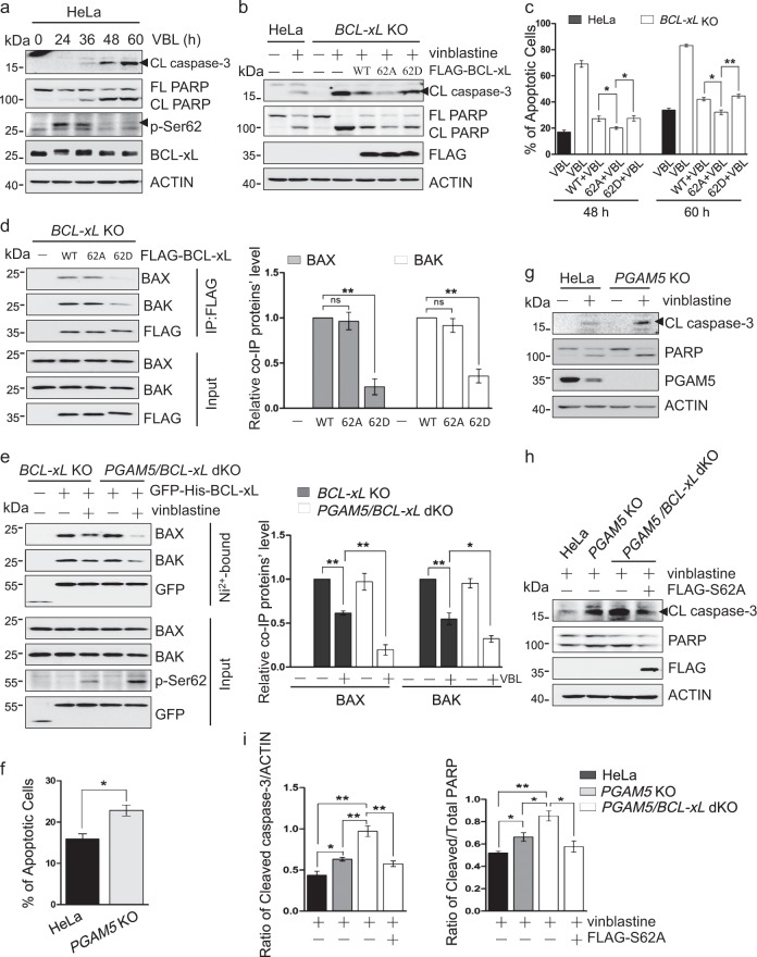
Fig. 2.
Dephosphorylation of BCL-xL by PGAM5 increases its anti-apoptotic activity. a HeLa cells were treated with vinblastine (VBL) for the indicated times, lysed and analyzed by western blotting with the indicated antibodies. CL caspase-3, cleaved caspase-3; FL PARP, full-length PARP; CL PARP, cleaved PARP. b HeLa or BCL-xL knockout HeLa cells with or without FLAG-BCL-xL, FLAG-BCL-xL-S62A, or FLAG-BCL-xL-S62D expression were treated with vinblastine for 48 h, lysed and analyzed by western blotting with the indicated antibodies. (c) HeLa or BCL-xL knockout HeLa cells expressing FLAG-BCL-xL or its mutants were treated with vinblastine for indicated times. After treatment, the cells were collected and stained with FITC-annexin V and propidium iodide (PI), then analyzed by flow cytometry. The percentages of apoptotic cells (annexin V+/PI− and annexin V+/PI+) were calculated. Data are the mean ± SEM of three independent experiments. **p < 0.01; *p < 0.05. d BCL-xL knockout HeLa cells were transfected with FLAG-tagged wild-type BCL-xL (WT) or the S62A or S62D mutant for 24 h, lysed and immunoprecipitated with anti-FLAG antibody. The co-immunoprecipitated endogenous BAX and BAK proteins were detected by western blotting. Right: densitometric quantification of co-immunoprecipitated BAX or BAK. Normalized to the bands from the wild type FLAG-BCL-xL expressing samples. Data are the mean ± SEM of three experiments. **p < 0.01. e BCL-xL or PGAM5/BCL-xL knockout HeLa cells were transfected with GFP-His-BCL-xL for 24 h, treated with vinblastine for 24 h, and lysed. The GFP-His-BCL-xL protein was isolated using Ni2+-chelating beads, eluted, and analyzed by western blotting with the indicated antibodies to detect the GFP-His-BCL-xL and the co-isolated BAX and BAK. Right: densitometric quantification of co-immunoprecipitated BAX or BAK. Normalized to the bands from the GFP-His-BCL-xL expressing untreated BCL-xL knockout HeLa cells samples. Data are the mean ± SEM of three experiments. *p < 0.05; **p < 0.01. f Wild type or PGAM5 knockout HeLa cells were treated with 500 nM vinblastine for 48 h, then stained with annexin V and propidium iodide (PI), and analyzed by flow cytometry. Data are the mean ± SEM of three experiments. *p < 0.05. g HeLa or PGAM5 knockout HeLa cells were treated without or with 500 nM vinblastine for 48 h, lysed and analyzed by western blotting with the indicated antibodies. h HeLa, PGAM5 knockout or PGAM5/BCL-xL double knockout cells, transfected with FLAG-BCL-xL-S62A if indicated, were treated with vinblastine for 48 h, lysed and analyzed by western blotting with the indicated antibodies. i The ratio of cleaved caspase-3 to ACTIN and the ratio of cleaved PARP to total PARP in (h) were calculated, respectively. Data are the mean ± SEM of three experiments. *p < 0.05; **p < 0.01

