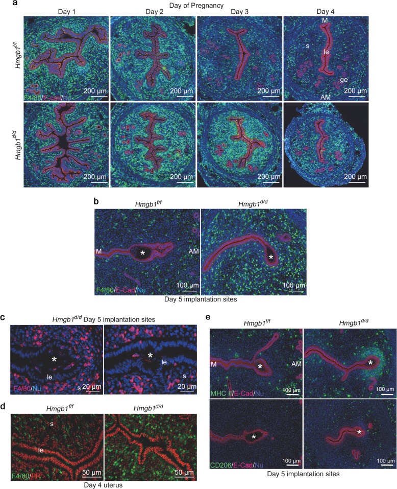
Fig. 5.
Deletion of Hmgb1 in uteri results in abnormal macrophage infiltration. a IF of F4/80 (macrophage marker) and E-Cad in days 1–4 pregnant uteri from Hmgb1f/f and Hmgb1d/d females. le luminal epithelium, s stroma, ge glandular epithelium, M mesometrial pole, AM antimesometrial pole. Scale bar: 200 μm. b IF of F4/80 and E-Cad in day 5 pregnant uteri of Hmgb1f/f and Hmgb1d/d females. Asterisks indicate the location of blastocysts. M mesometrial pole, AM antimesometrial pole. Scale bar: 100 μm. c IF of F4/80 at higher magnification of day 5 pregnant uterine sections from Hmgb1d/d females. Invasion of macrophages into the implantation chamber (crypt) was observed. Asterisks indicate the location of blastocysts. le luminal epithelium, s stroma, IS implantation site. Scale bar: 20 µm. d IF of F4/80 and PR in day 4 pregnant uteri from Hmgb1f/f and Hmgb1d/d females. le luminal epithelium, s stroma. Scale bar: 50 µm. e IF of MHC II (M1 macrophage marker; upper panels), CD206 (M2 macrophage marker; lower panels), and E-Cad in day 5 Hmgb1f/f and Hmgb1d/d pregnant uteri. Asterisks indicate the location of blastocysts. M mesometrial pole, AM antimesometrial pole. Scale bar: 100 µm. Each image is a representative from at least three independent experiments. All uteri were collected at 9–10 a.m. on the indicated day of pregnancy. See also Fig. S5, Movies S1 and S2

