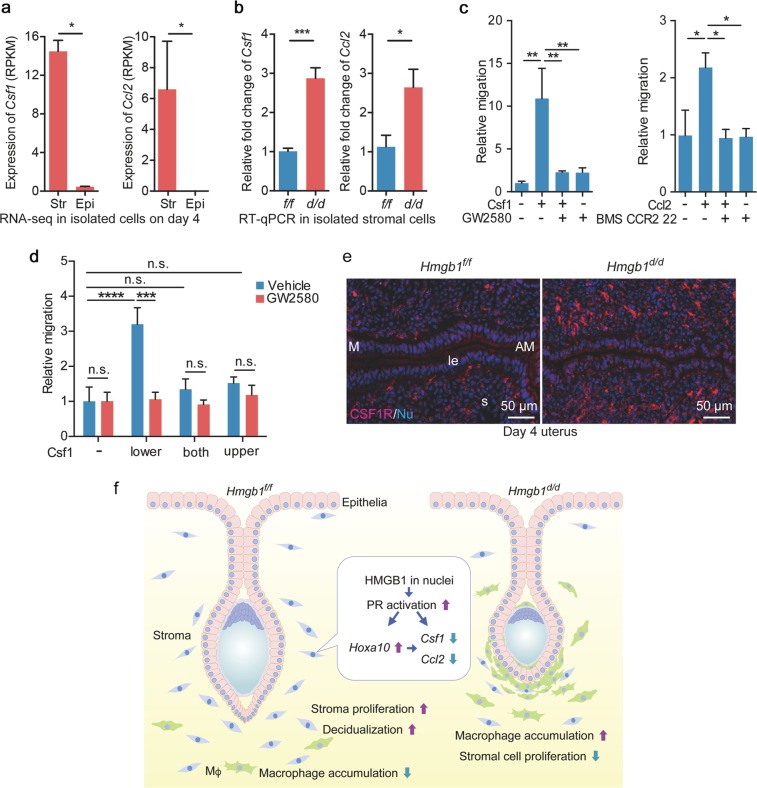
Fig. 6.
Hmgb1-deleted stromal cells cause increased cytokine levels. a Expression levels (RPKM) of macrophage attractants, Csf1 and Ccl2, were analyzed by RNA-seq analysis in isolated epithelial/stromal cells from day 4 uteri. n = 3 for each group. Str stroma, Epi epithelium. Data are presented as mean ± SEM, *P < 0.05 by two-tailed Student’s t -test. b Quantitative RT-PCR showed increased levels of macrophage attractants (Ccl2 and Csf1). n = 4 for each genotype. Data are presented as mean ± SEM, *P < 0.05 and ***P < 0.001 by two-tailed student’s t test. c Migration assays show that Csf1 and Ccl2 attract macrophages depending on their specific receptors. GW2580 and BMS CCR2 22 were used as Csf1 and Ccl2 receptor antagonists, respectively. These assays were performed in three wells for each group and three fields/wells were quantified. Data are presented as mean ± SEM, *P < 0.05 and **P < 0.01 (one-way ANOVA followed by Bonferroni’s post hoc test). d Migration assays show that Csf1 in the lower chamber attracts macrophages seeded in the upper chamber depending on its specific receptor. The assays were performed in three wells for each group and three fields/wells were quantified. Data are presented as mean ± SEM, *P < 0.05 and **P < 0.01 (two-way ANOVA followed by Bonferroni’s post hoc test). e IF of CSF1R in day 4 pregnant uteri from Hmgb1f/f and Hmgb1d/d females. le luminal epithelium, s: stroma, M mesometrial pole, AM antimesometrial pole. Scale bar: 50 µm. Each image is a representative from at least three independent experiments. f Schematic diagram showing the function of HMGB1 in uteri during pregnancy. All uteri were collected at 9–10 a.m. on day 4 of pregnancy

