Fig. 3.
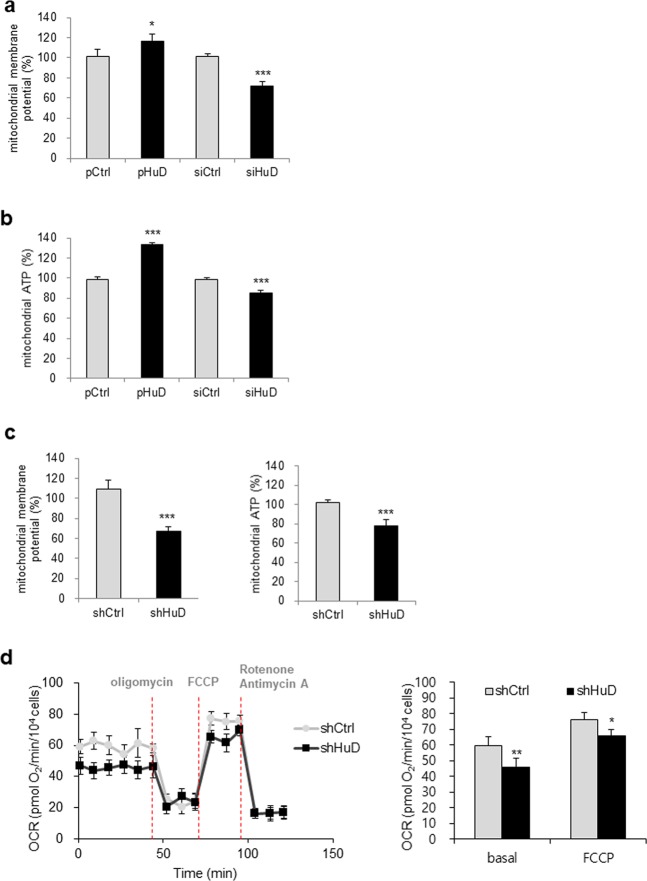
Regulation of mitochondrial activity by HuD. a, b After transfection of βTC6 cells with HuD siRNA or pHuD with appropriate controls, mitochondrial membrane potential (a) and mitochondrial ATP levels (b) were analyzed. Cells were stained with tetraethyl benzimidazoly carbocyanine iodide (JC-1) and the fluorescence was measured at 530 nm (excitation)/590 nm (emission). Cells pretreated with galactose-containing media were incubated with the ATP detection reagent and the luminescence was measured using a microplate reader. c Mitochondrial membrane potential and ATP level were determined in βTC6-shHuD and βTC6-shCtrl cells. d Mitochondrial respiration in βTC6-shCtrl and βTC6-shHuD cells was determined by assessing the oxygen consumption rate. Cells were sequentially incubated with oligomycin, FCCP, rotenone, and antimycin A and OCR was measured using the Seahorse FX24 Extracellular Flux Analyzer. Data represent the mean ± SEM derived from three independent experiments. *p < 0.05; **p < 0.01; ***p < 0.001

