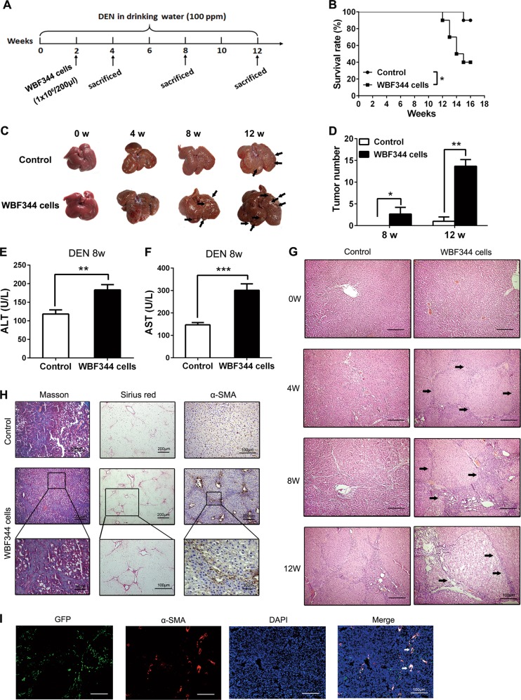
Fig. 2.
Hepatic progenitor cell (HPC) transplantation promoted hepatocarcinogenesis and liver fibrosis in primary liver cancer model. a F344 rats were intrasplenically injected with HPC-like WB-F344 cells (1 × 10⁶/200 μl) at 2 weeks after diethylnitrosamine (DEN) treatment and sacrificed at different time points to observe the development of hepatocellular carcinoma (HCC). b DEN-treated F344 rats injected with WB-F344 cells; survival rate was observed. Data are represented as mean ± SD. *p < 0.05. c, d Rat livers of the indicated groups. The number of HCC nodules per liver in rats was determined at 8 and 12 weeks after DEN treatment. Data are represented as mean ± SD. *p < 0.05, **p<0.01. e, f Serum levels of aspartate aminotransferase (AST) and alanine aminotransferase (ALT) were determined to indicate the extent of liver damage caused by DEN administration and the role of HPCs in promoting the damage. Data are represented as mean ± SD. **p < 0.01, ***p<0.001. g Hematoxylin and eosin (H&E) staining was employed to observe the histological structure in different groups. h Hepatic collagen deposition was determined by Sirius Red staining and Masson’s trichrome staining. The expression of α-smooth muscle actin (α-SMA) was detected by immunohistochemical analysis. i WB-F344 cells were transfected with GFP-labeled lentiviral vector. After transplantation in rats, WB-F344 cells in the liver were detected by immunofluorescence double staining of GFP and α-SMA expression. The merged immunofluorescence staining picture was used to determine the expression of α-SMA in GFP-positive cells

