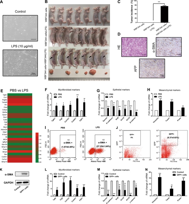
Fig. 5.
Lipopolysaccharide (LPS) induced hepatic progenitor cell (HPC) differentiation into myofibroblasts, which enhanced the tumorigenic potential of HPCs. a Cell morphology was observed in LPS-disposed WB-F344 cells for 2 weeks. b, c Tumorigenicity was detected in each group. Pictures were taken at the eighth week after subcutaneous injection. d Subcutaneous tumor was observed with hematoxylin and eosin (H&E) staining. α-Fetoprotein (AFP) and α-smooth muscle actin (α-SMA) expression was detected by immunohistochemical analysis. e RNA was extracted from WB-F344 cells after 2 weeks of LPS exposure and subjected to microarray analysis (n = 3 for each sample). Heatmap displaying positive fold changes in expression of myofibroblast-like genes in phosphate-buffered saline (PBS)-treated WB-F344 cells versus LPS-treated WB-F344 cells. f Expression of myofibroblast-associated genes was examined by real-time PCR (n = 3; mean ± SD). *p < 0.05, **p < 0.01, and ***p < 0.001. g, h Expression of epithelial and mesenchymal markers were examined by real-time PCR (n = 3; mean ± SD). **p < 0.01 and ***p < 0.001. i Flow cytometry was performed to analyze α-SMA expression in PBS-treated WB-F344 cells versus LPS-treated WB-F344 cells (n = 3; mean ± SD). j WB-F344 cells were transfected with GFP-labeled lentiviral vector. Then, F344 rats were intrasplenically injected with GFP-labeled WB-F344 cells at 2 weeks after DEN treatment. After transplantation in DEN-induced rats for 6 weeks, GFP-positive cells were isolated from the liver by using flow cytometry. k. Western blot was employed to detect α-SMA expression in GFP-positive cells and WB-F344 cells. l Expression of myofibroblast-associated genes in GFP-positive cells and WB-F344 cells was examined by real-time PCR (n = 3; mean ± SD). **p < 0.01 and ***p < 0.001. m, n Expression of epithelial and mesenchymal markers in GFP-positive cells and WB-F344 cells were examined by real-time PCR (n = 3; mean ± SD). *p < 0.05, **p < 0.01, and ***p < 0.001

