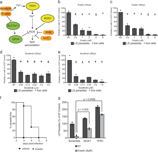
Fig. 4.
Induction of ferroptosis-like signaling with small molecules eliminates Plasmodium LS parasites in vitro and in vivo. a Signaling downstream of Erastin treatment as reported in the literature. b, c Hepa1-6 cells were infected with 5 × 10⁴ P. yoelii sporozoites and treated with Erastin at indicated concentrations 90 min after infection. After b 24 h or c 48 h, LS parasites were visualized by Py HSP70 staining and quantified by fluorescent microscopy. Cell death in uninfected cells was quantified by Trypan Blue staining. d, e Hepa1-6 cells were infected with P. yoelii and treated with Sorafenib at indicated concentrations. After 24 h (d) or 48 h (e), LS parasites were visualized by PyHSP70 staining and quantified by fluorescent microscopy. Cell death in uninfected cells was evaluated by Trypan Blue staining. f 10 C57Bl/6 mice were treated with 30 mg/kg Erastin or vehicle control for 4 days. On the second day of treatment, mice were challenged with 1000 P. yoelii sporozoites by retro-orbital injection. Beginning on day 3 post-infection, blood was evaluated by giemsa-stained thin smear for the presence of blood stage parasites. Each bar represents the mean of replicates. g 1.5 × 10⁵ Hepa1-6 cells transduced with lentivirus expressing shRNAs against a scramble control, NOX1 or TFR1 and infected with 5 × 10⁴ P. yoelii sporozoites. After 90 min post-infection, cells were treated with 8 µM Erastin or a DMSO control. Parasites were visualized 24 h post-infection by Hsp70 staining and quantified by microscopy. Each bar represents the mean of replicates. P-values were obtained using a Student’s t-test. In a–e and g, points represent individual technical replicates and are representative of three independent experiments

