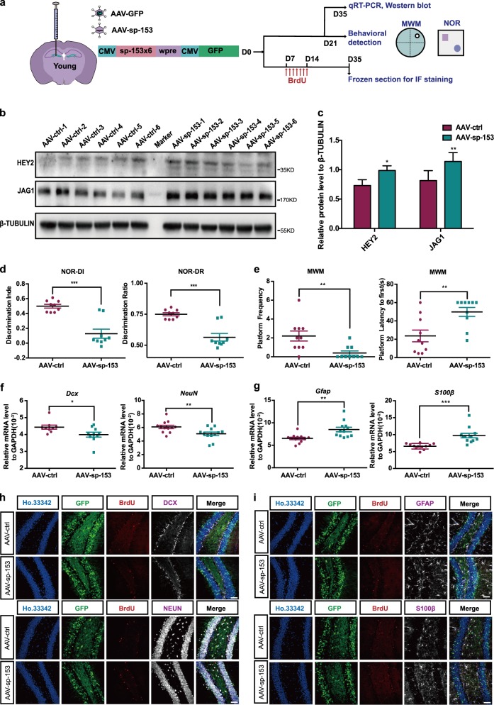
Fig. 6.
Inhibition of miR-153 decreases hippocampal neurogenesis and impairs the cognitive functions of young mice. a Schematic of virus injection into the hippocampi of young mice and diagram of the detection process. MWM Morris water maze, NOR novel object recognition. b Western blot analysis of the protein levels of JAG1 and HEY2 in the hippocampi of AAV-ctrl and AAV-sp-153 young mice normalized to β-TUBULIN. c Statistical analysis of the grayscale levels of JAG1 and HEY2 by ImageJ; AAV-ctrl, n = 6; AAV-sp-153, n = 6. d Discrimination index and discrimination ratio of novel objects; AAV-ctrl, n = 10; AAV-sp-153, n = 9. e Platform crossings and amounts of time required for the mice to first reach the platform area during the water maze test; AAV-ctrl, n = 10; AAV-sp-153, n = 10. f, g Quantitative RT-PCR detection of the newborn neuronal marker Dcx (AAV-ctrl, n = 9; AAV-sp-153, n = 10), the mature neuronal marker NeuN (AAV-ctrl, n = 12; AAV-sp-153, n = 12), and the glial cell markers Gfap (AAV-ctrl, n = 11; AAV-sp-153, n = 11) and S100β (AAV-ctrl, n = 12; AAV-sp-153, n = 12) in the hippocampi of AAV-ctrl and AAV-sp-153 young mice. h Immunofluorescence analysis of neurons in the hippocampi of AAV-ctrl and AAV-sp-153 young mice as evaluated by GFP (green), BrdU (red), DCX (white), NEUN (white), and Ho.33342 (blue). Scale bar: 50μm. i Immunofluorescence analysis of astrocytes in the hippocampi of AAV-ctrl and AAV-sp-153 mice as evaluated by GFP (green), BrdU (red), GFAP (white), S100β (white), and Ho.33342 (blue). Scale bar: 50 μm. Data information: the data shown are the mean ± SEM; Student’s t test; ***p < 0.001, **p < 0.01, *p < 0.05. AAV-sp-153 AAV-sponge-153, young mice 8–10 weeks old

