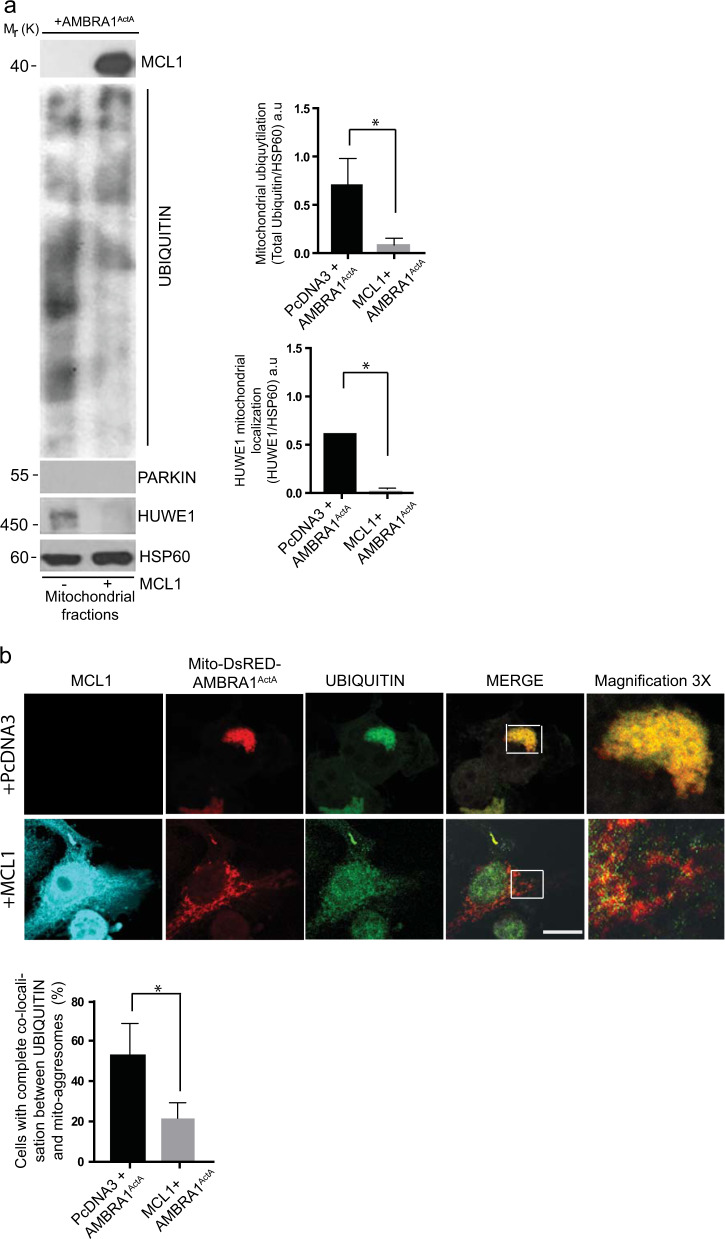
Fig. 2.
Upon AMBRA1-mediated mitophagy, MCL1 inhibits HUWE1 translocation to mitochondria and their ubiquitylation. a HeLa cells were cotransfected with Myc-AMBRA1ActA and PcDNA3 or MCL1. After 24 h of transfection, mitochondrial enrichment was performed through differential centrifugations, mitochondrial fractions were analyzed using the indicated antibodies and anti-HSP60 was used as a loading control. The graphs illustrate UBIQUITIN/HSP60 and HUWE1/HSP60 ratio (±S.D.) as an arbitrary unit (a.u). Each point value represents the mean ± S.D. from three independent experiments. Statistical analysis was performed using Student’s test (*P < 0.05). b HeLa cells were cotransfected with a vector encoding Mito-DsRED-AMBRA1ActA and PcDNA3 or MCL1. Following 24 h of transfection, cells were fixed and assessed by immunolabelling using anti-MCL1 and UBIQUITIN antibodies. Nuclei were stained with DAPI 1 µg/µl 20 min. The merging of the fluorescence signals is illustrated. Scale bar: 8 µm. The graph illustrates the percentage of cells showing a complete colocalization between UBIQUITIN and the mitochondrial network (±S.D). Each point value represents the mean ± S.D. from three independent experiments. Statistical analysis was performed using Student’s test (*P < 0.05)

