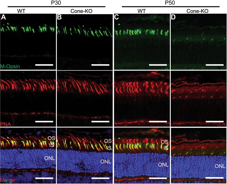
Fig. 6.
Loss of Emc3 in mammalian cone cells led to cone cell death. a, b Retina cryosections from (a) WT and (b) cone-KO littermate mice at P30. Sections were immunostained for M-Opsin (green) and the cone marker, peanut agglutinin (PNA, red). Cone cell number did not decrease at P30. c, d Retina cryosections from (c) WT and (d) cone-KO mice at P50. Sections were immunostained for M-Opsin (green) and PNA (red). There were almost no cone cells in cone-KO retinas. Nuclei were counterstained with DAPI. ONL outer nuclear layer, OS outer segment, IS inner segment. Scale bar is 25 μm

