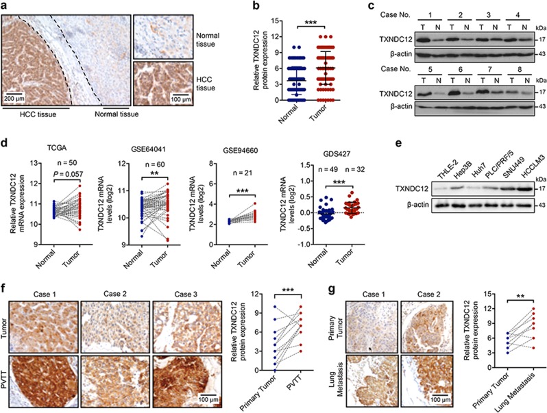
Fig. 1.
TXNDC12 is upregulated in human hepatocellular carcinoma. a Representative immunohistochemical staining of TXNDC12 in human hepatocellular carcinoma (HCC) tissues; scale bars: 40 μm (left), 20 μm (right). b Quantification of TXNDC12 expression in human HCC tissues (n = 106). The data are the means ± SDs. c Representative images of TXNDC12 expression obtained by immunoblot analysis in HCC tissues. d Relative mRNA expression of TXNDC12 in the TCGA dataset and three GEO datasets (GSE64041, GSE94660, and GDS427). The data are the means ± SDs. e Relative protein expression of TXNDC12 in normal hepatocytes and four HCC cell lines, as obtained by immunoblot analysis. f Representative images and quantification of TXNDC12 expression in portal vein tumor thrombus and matched primary HCC tissues obtained by immunohistochemical analysis (n = 14); scale bar: 20 μm. g Representative images and quantification of TXNDC12 expression in lung metastases and matched primary HCC tissues obtained by immunohistochemical analysis (n = 10); scale bar: 20 μm. **P < 0.01; ***P < 0.001

