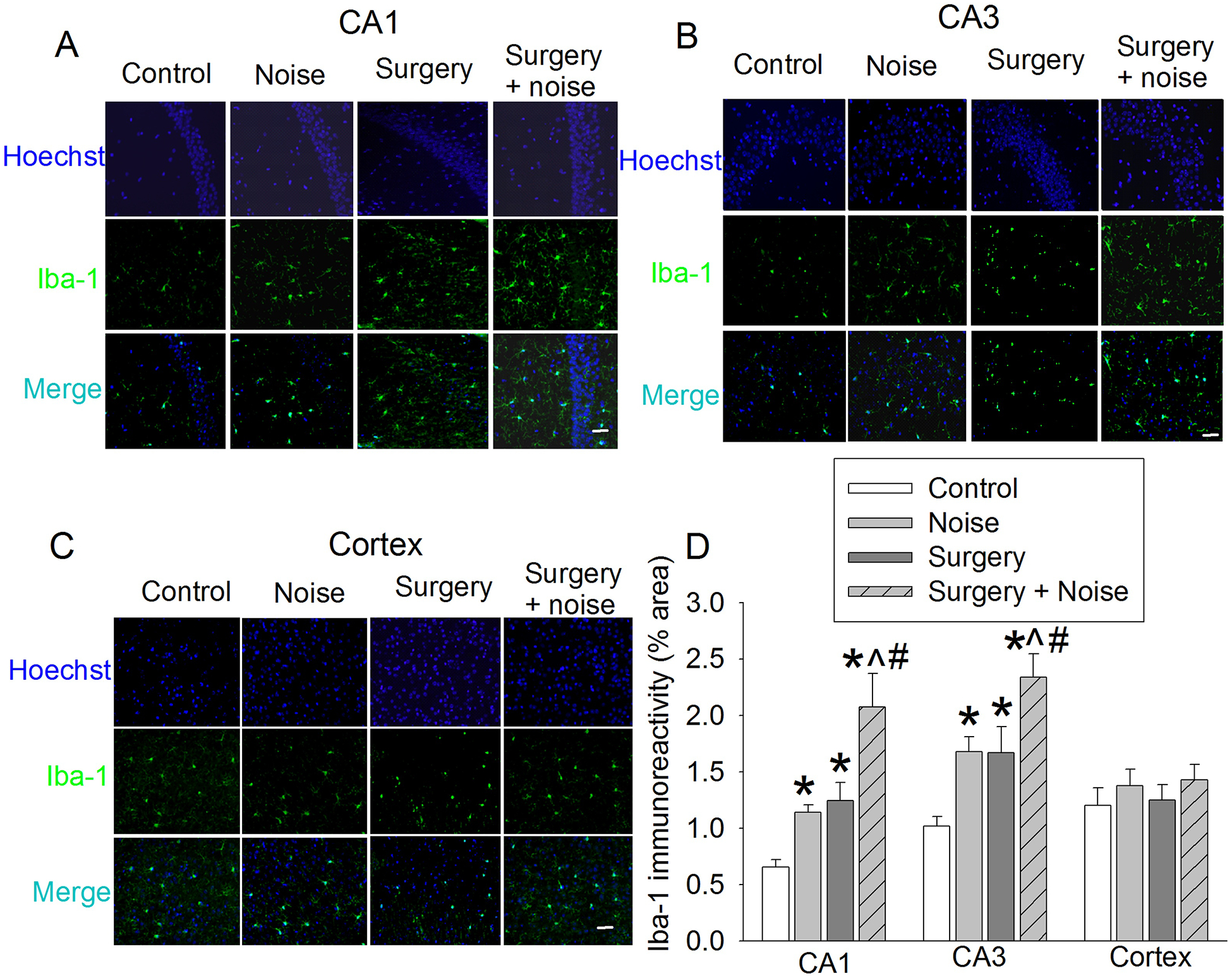
Fig. 3. Effects of noisy environment and surgery on Iba-1 expression in the brain.

Seven-week old mice were subjected to right carotid exploration and 75 db noisy environments. Hippocampus and cortex were harvested at 6 h after surgery or the last episode of noise. A: Representative immunostaining images of Iba-1 in CA1. B: Representative immunostaining images of Iba-1 in CA3. C: Representative immunostaining images of Iba-1 in cerebral cortex. D: Graphic presentation of the percentage area that is Iba-1-postive staining in CA1, CA3, and cerebral cortex. Scale bar = 30 μm. Results are means ± S.E.M. (n = 6). * P < 0.05 compared with control, ^ P < 0.05 compared with noise group, # P < 0.05 compared with surgery group.
