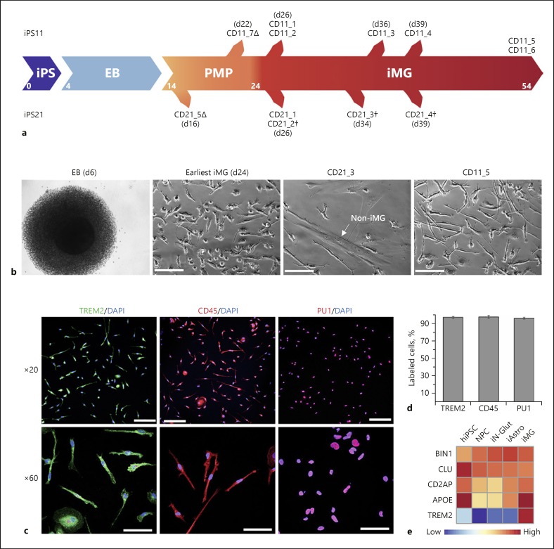
Fig. 1.
Generation of hiPSC-derived microglia (iMG). a hiPSC lines iPS11 and iPS21 are differentiated to microglia lines CD11 and CD21, respectively, via embryoid bodies (EB) and primitive macrophage precursors (PMP) over a period of 24 days. PMPs are produced continuously in culture and are terminally differentiated into microglia when required. CD21 and CD11 samples with a delta (∆) denote iPMP samples and a dagger (†) denotes mixed samples containing non-iMG cells. b DIC images of EB at day 6 (far left), iMG cells at day 24 (near left, CD11_5), and more mature microglia cells at day 44 (far right, CD11_5). Some of the iMG cultures also had non-iMG cells (near right, CD21_3), which morphologically resembled fibroblasts. Scale bars, 500 µm. c Immunofluorescence staining of iMG from line CD11 at day 24 shows expression of microglial/macrophage markers TREM2, CD45, and PU1. Scale bars in ×20 images, 100 µm; scale bars in ×60 images, 50 µm. d The majority of the iMG cells from line CD11 show expression of microglial/macrophage markers TREM2, CD45, and PU1 (n = 4–5). e Expression by qPCR of AD risk genes (BIN1, CLU, CD2AP, APOE, TREM2) in different cell types (normalized to GAPDH). iAstro, hiPSC-derived astrocytes; iN-Glut, hiPSC-derived glutaminergic neurons; NPC, neural progenitor cells.

