Fig. 7.
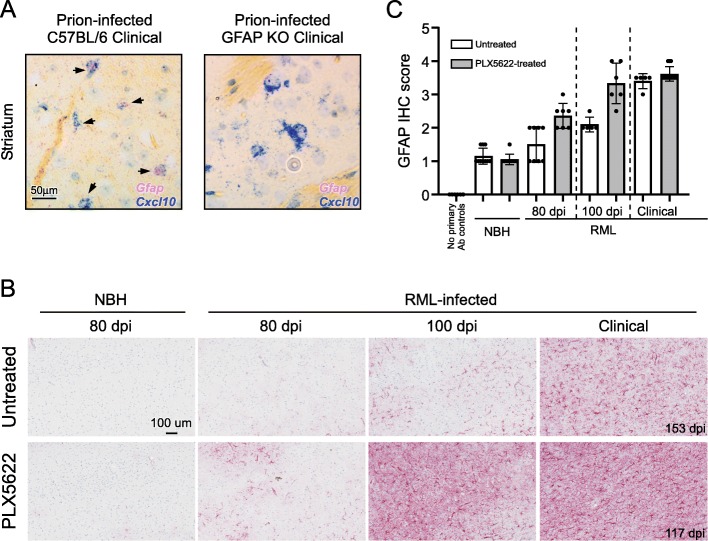
Representative dual colorimetric in situ hybridization (CISH) to determine cellular population expressing Cxcl10 (a). Prion-infected C57BL/6 and GFAP-deficient (GFAP KO) mice were inoculated with prions and allowed to advance to clinical disease. Sections from formalin fixed brains were hybridized with probes specific for Cxcl10 (blue) or Gfap (pinkish-red). Infected C57BL/6 mice had several large nuclei demonstrating expression of both Gfap and Cxcl10 (arrows) within the cell in several regions including the cerebral cortex (not shown), cerebellum, (not shown) and striatum (shown). GFAP KO mice displayed many cells in these same regions that expressed Cxcl10 that lacked hybridization with the Gfap probe, demonstrating the hybridization with Gfap was specific. The scale for both representative images is indicated in the first image in A. Representative immunohistochemical assessment of astrogliosis in cerebral cortex brain sections from Untreated and PLX5622-treated mice (b). Mice were inoculated with either normal brain homogenate (NBH) or scrapie strain RML. Sections of cerebral cortex from mice at the dpi indicated were probed with antibodies against GFAP. Representative images of the cerebral cortex are shown for all at the scale indicated in the first panel. The dpi of the clinical mice is as indicated. The intensity of GFAP histopathological immunoreactivity observed from stained coronal sections (Additional File, Figure S1) was given a subjective score from 0 to 4 based on the amount and distribution of GFAP staining in the brain (see methods) and is graphically depicted (c). No primary antibody (Ab) controls were absent of staining and defined as 0

