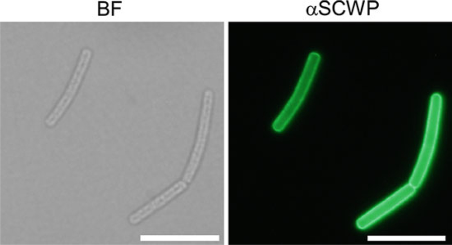
Fig. 1.

Micrographs of B. anthracis strain Sterne cells revealing the surface display of SCWP. Cells were fixed in formalin, stained with anti-SCWP antibodies (αSCSWP) and observed by bright-field microscopy (BF, left panel) or fluorescence microscopy (right panel). Scale bar, 10 μm
