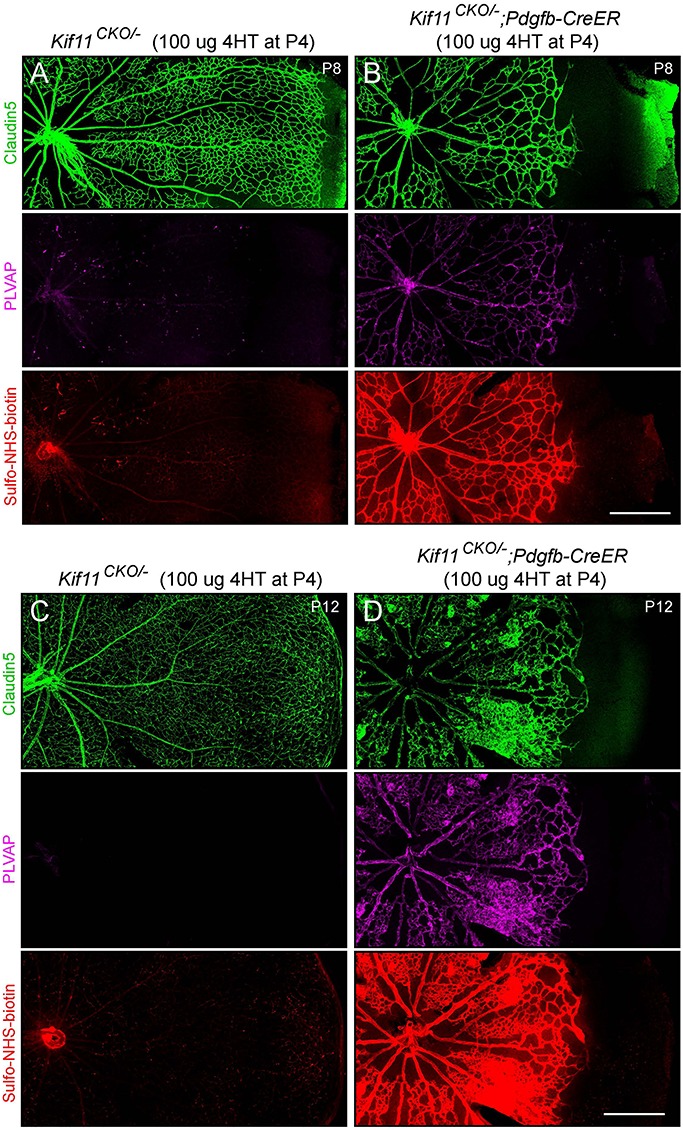
Figure 2.

Severely retarded retinal vascular growth following the early postnatal EC-specific knockout of Kif11. Retinal flatmounts were prepared at P8 or P12 from littermate control Kif11CKO/− mice (A,C) and Kif11CKO/−;Pdgfb-CreER mice (B,D) following the treatment with 100 μg 4HT at P4. Each image shows one retinal quadrant, with the optic disc at the left and the retinal periphery at the right. Loss of Kif11 leads to retarded vascular development and reduced vascular coverage of the retinal surface. At both ages, Kif11CKO/−;Pdgfb-CreER retinas show increased expression of PLVAP and increased Sulfo-NHS-biotin leakage. Scale bars, 500 μm.
