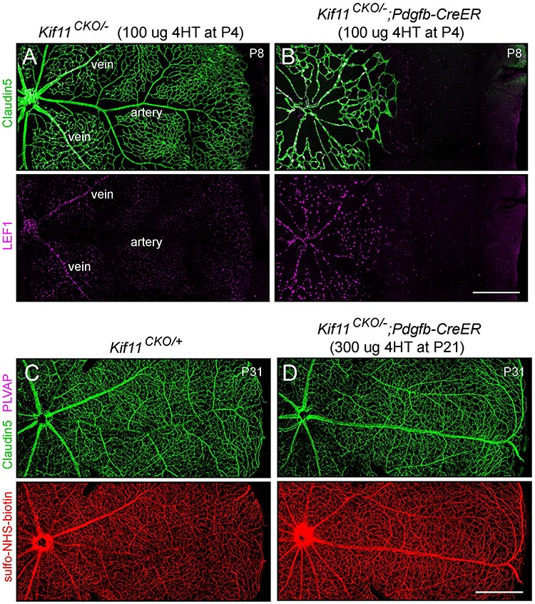
Figure 3.

EC-specific knockout of Kif11 leads to continued EC accumulation of LEF1 during development and no effect of post-mitotic knockout on vascular permeability. (A,B) Retinal flatmounts were prepared at P8 from littermate control Kif11CKO/− mice (A) and Kif11CKO/−;Pdgfb-CreER mice (B) following the treatment with 100 μg 4HT at P4. Each image shows one retinal quadrant, with the optic disc at the left and the retinal periphery at the right. LEF1, a marker of beta-catenin signaling, accumulates in the nuclei of vein and capillary ECs but not in arterial ECs in control retinas and in all ECs in mutant retinas. (C,D) Retinal flatmounts were prepared at P31 from littermate control Kif11CKO/+ mice (C) and Kif11CKO/−;Pdgfb-CreER mice (D) following the treatment with 300 μg 4HT at P21. Each image shows one retinal quadrant as in (A) and (B). Loss of Kif11 has no effect on vascular architecture, expression of Claudin5, suppression of PLVAP or BRB integrity, as seen by the absence of Sulfo-NHS-biotin leakage from the intravascular space into the retinal parenchyma. See Supplementary Material Figure S1 for an analysis of Cre-mediated recombination at P21. Scale bars, 500 μm.
