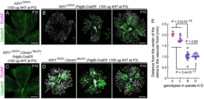
Figure 4.

Postnatal EC-specific stabilization of beta-catenin does not rescue the Kif11 retinal vascular phenotype. (A,B) Retinal flatmounts were prepared at P10 from control Kif11CKO/+ mice (A) and Kif11CKO/−;Pdgfb-CreER mice (B; three examples shown) following the treatment with 100 μg 4HT at P3 to assess the Kif11 retinal vascular phenotype. (C,D) From the same set of crosses, retinal flatmounts were prepared at P10 from control Kif11CKO/+;Ctnnb1flex3/+;Pdgfb-CreER mice (C) and Kif11CKO/−;Ctnnb1flex3/+;Pdgfb-CreER mice (D; three examples shown) following the treatment with 100 μg 4HT at P3. Retinal vascular development in both sets of control mice is indistinguishable from WT. Beta-catenin stabilization following the Cre-mediated recombination of Ctnnb1flex3/+ fails to rescue the Kif11 retinal vascular phenotype. In (A) and (C), each image shows one retinal quadrant, with the optic disc at the bottom and the retinal periphery at the top; in (B) and (D), each image is centered on the optic disc. All images in (A–D) are at the same magnification. Scale bars, 500 μm. (E) Quantification of the radial distance from the optic disc to the vascular front at P10 for individual retinal quadrants for the genotypes shown in panels A–D. Plots show mean ± standard deviation.
