Figure 5.

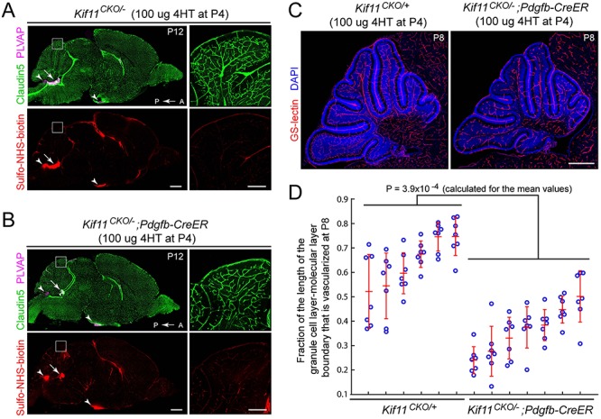
Retarded density of cerebellar vasculature following the early postnatal EC-specific knockout of Kif11. (A,B) Sagittal brain sections were prepared at P12 from control Kif11CKO/− mice (A) and Kif11CKO/−;Pdgfb-CreER mice (B) following the treatment with 100 μg 4HT at P4. The region of the dorsal cerebellum enclosed by the white square is enlarged at right. Scale bars, 1 mm (left images) and 200 μm (right images). Except for modestly reduced vascular density in the cerebellum (inset at right), brain size and vascular density are unaffected by EC-specific loss of Kif11. Sulfo-NHS-biotin leakage is seen in the choroid plexus (arrow) and in the circumventricular organs (area postrema and median eminence; arrowheads) as expected but is not observed elsewhere in the brain. (C) Sagittal brain sections were prepared at P8 from control Kif11CKO/+ mice and Kif11CKO/−;Pdgfb-CreER mice, following the treatment with 100 μg 4HT at P4. Scale bar, 500 μm. (D) With early postnatal loss of Kif11, vascular density in the P8 cerebellum is reduced, as determined by quantifying the fraction of the molecular layer/granule cell layer border that is occupied by blood vessels in six control Kif11CKO/+ and seven Kif11CKO/−;Pdgfb-CreER P8 mice. For each mouse, seven folia were quantified from a single confocal stack of the cerebellum [examples in (C)]. The plot in (D) shows mean ± standard deviation for each mouse.
