
Figure 1.
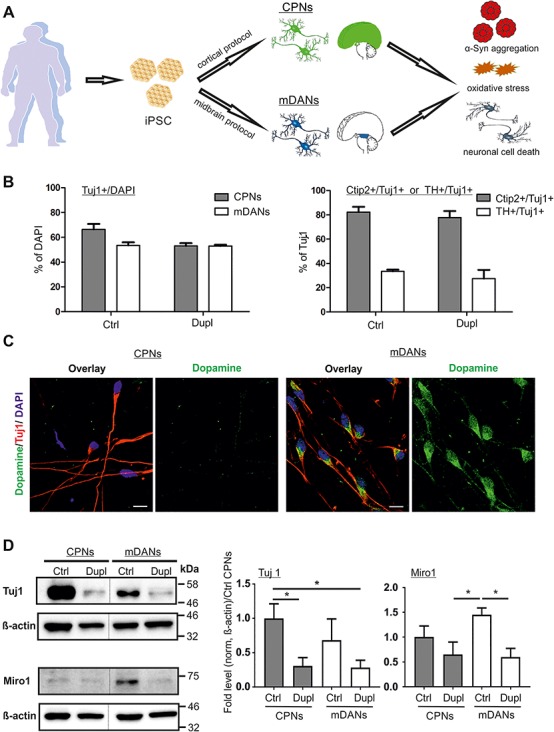
α-Syn gene (SNCA) locus duplication (Dupl) does not alter neuronal differentiation efficiency. (A) Schematic summary of the study. CPNs and mDANs were differentiated from human iPSCs of a control individual (Ctrl) and PD patients carrying SNCA Dupl (Dupl) using cortical and small-molecule-based midbrain protocols, respectively. α-Syn aggregation was analyzed biochemically. Oxidative stress was evaluated by the level of ROS and protein nitration. Neuronal cell death was determined by ICC analysis of cleaved Caspase-3 (C-Casp3). (B) Neuronal differentiation was equally effective in Ctrl and Dupl as assessed by ICC of neuronal marker β3-tubulin (Tuj1), cortical projection marker Ctip2 and midbrain dopaminergic marker TH (representative images used for the quantification are shown in Fig. 4A). Amounts of neurons were determined as the percentage (%) of Tuj1-positive cells over DAPI-positive cells, whereas cortical and midbrain neurons were evaluated as the proportion (%) of Ctip2- or TH-positive neurons over total Tuj1-positive neurons in three independent differentiation rounds. Values are shown as mean ± SD. Two-tailed Student’s t-test was used. (C) ICC staining of dopamine in iPSC neuronal cultures revealed positive dopamine signals in Tuj1-positive neurons, differentiated from Ctrl iPSCs by the midbrain protocol, while dopamine was barely detectable in Ctrl CPNs, confirming the neurotransmitter-specific phenotype of mDANs. Scale bars 10 μm. (D) Tuj1 and Miro1 protein expression was assessed in Ctrl and PD Dupl CPNs and mDANs by WB. Quantification was conducted by the normalization of Tuj1 or Miro1 signals to β-actin levels followed by setting Ctrl CPN levels to 1. Reduced Tuj1 and Miro1 expression was determined in both PD Dupl CPNs and mDANs compared with Ctrl neurons. *P ≤ 0.05; values are shown as mean ± SD (three independent experiments). One-way ANOVA with multiple comparisons test was used. Blots for Tuj1 and Miro1 within one black frame are derived from the same membrane. β-Actin was used as a loading control and was probed on the same membrane as the respective target protein. Lanes from different parts of the same membrane are separated by black dashed lines.