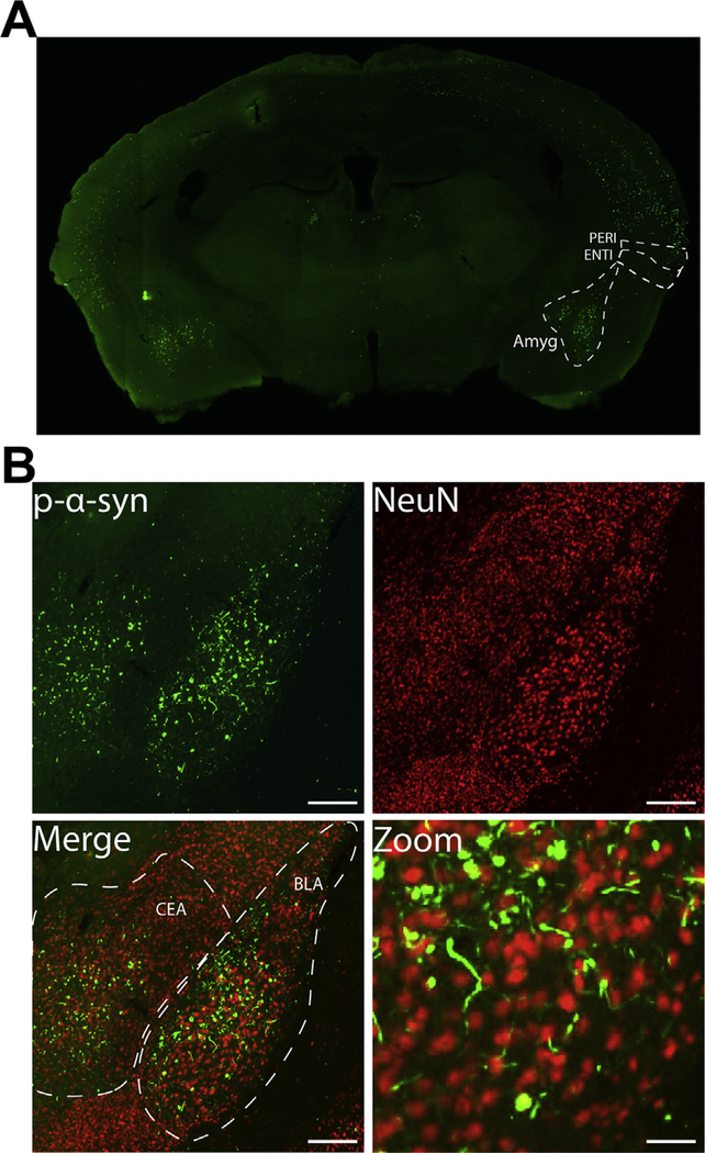
Fig. 1.
Bilateral fibril injection leads to α-synuclein inclusion formation in the basolateral and central amygdala. Mice received bilateral injections of α-syn fibrils at 3–4 months of age. A) pSer129-α-synuclein (EP1536Y) immunofluorescence from a representative coronal section showing inclusions in the basolateral and central amygdala. B) Higher magnification of immunofluorescence for pSer129-α-synuclein (green) and NeuN (red) showing inclusions in neurons in the basolateral and central nuclei of the amygdala. Scale bar = 200 μm and 50 μm (zoom). Abbreviations: Amyg = amygdala, BLA = basolateral nucleus of the amygdala, CEA = central nucleus of the amygdala, ENTI = entorhinal cortex, PERI = perirhinal cortex. (For interpretation of the references to colour in this figure legend, the reader is referred to the web version of this article.)

