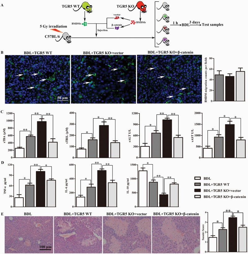
Fig. 7.
Administration of TGR5-deficient BMDMs aggravated BDL-induced CHI via modulating β-catenin signaling. (A) BMDMs isolated from WT and TGR5−/− mice were cultured and differentiated for 7 days. TGR5−/− BMDMs differentiated under L929 conditions were transfected with β-catenin–over-expressing or control vector, and 5 × 10⁶ cells were injected into the tail veins of irradiated mice. Bone marrow-irradiated C57BL/6 mice were subjected to BDL at 1 h after injection. Untreated BDL-induced CHI immune-deficient mice were used as controls. Mice were sacrificed at 3 days after BDL. (B) The cell membrane of each group of cells was labeled with PKH67 in vitro, and then cells were injected from the tail vein. The migration of injected macrophages into liver of the recipient mice was determined by tissue section staining (white arrows, scale bars: 50 μm). (C) Hepatocellular function was evaluated by sTBA, sTBIL, sALT and sAST (n = 6 for each group). (D) Protein levels of TNF-α, IL-6 and IL-10 in serum measured by ELISA (n = 6). (E) Histopathologic analysis of livers harvested 3 days after BDL (n = 5), and severity of liver injury according to Suzuki’s histological grading. Scale bars: 100 μm. Statistical analysis was performed by Student’s t-test. The data were displayed as mean ± SD. *P < 0.05, **P < 0.01.

