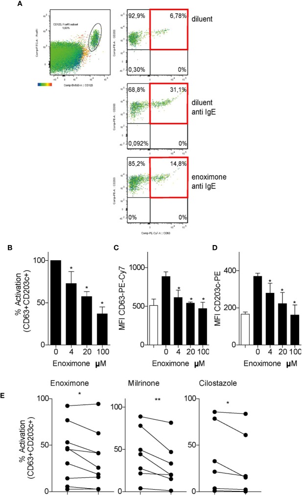
Figure 3.
PDE3 inhibitors reduce FcεR-mediated basophil activation. (A) Flow cytometry (FACS) plot of CD63/CD203 profiles of the CD123+FcεR+ basophil cell fractions from PBMCs that were treated with enoximone or diluent and stimulated with phosphate buffered saline (PBS) or anti-immunoglobulin E (IgE), as indicated. (B–D) Dose-response to enoximone for proportions of CD63+CD203+ activated cells within the fraction of CD123+FcεR+ PBMC (B), MFI values for CD63 (C), and MFI values for CD203c (D). Values are mean ± S.E.M; n=3. (E) Capacity of the PDE3i enoximone, milrinone and cilostazol to inhibit activation of basophils, shown as the proportions of CD63+CD203+ activated cells within the fraction of CD123+FcεR+ PBMC. A Wilcox signed test and a Mann-Whitney U test was used. *P < 0.05; **P < 0.01. Data are shown of one representative experiment from three independent experiments. MFI, median fluorescence intensity.

