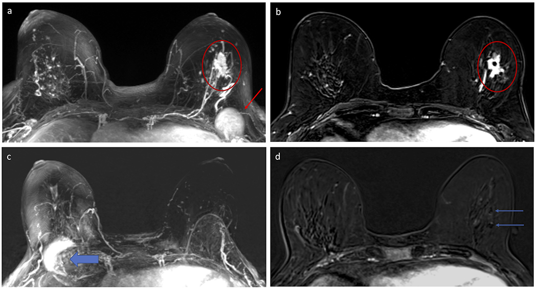
Figure 6:

Example of decrease in background parenchymal enhancement (BPE) in a 59 year-old woman who underwent neoadjuvant chemotherapy (NAC) for a poorly differentiated invasive ductal carcinoma (IDC) in the left breast. Subtracted maximum intensity projection (MIP) from the pre-NAC breast MRI (A) demonstrates moderate BPE in both breasts, with the left breast IDC (red circle) distinct from the BPE and an enlarged left level I axillary lymph node (red arrow) also evident. Selected post-contrast T1-weighted subtracted axial slice centered at the level of the left breast IDC demonstrates the mass with two clips within (red circle). Subtracted MIP from the post-NAC MRI performed five months later (B) demonstrates decrease in BPE, now minimal, with resolution of both the mass and axillary lymphadenopathy (note artifact on the MIP from the port-a-catheter in the right chest for chemotherapy, wide blue arrow). Patient was confirmed to have pathologic complete response (pCR) on surgery. Several papers have suggested that a reduction in BPE after NAC could help identify which patients have better clinical outcomes, including pCR.
