
Figure 7:
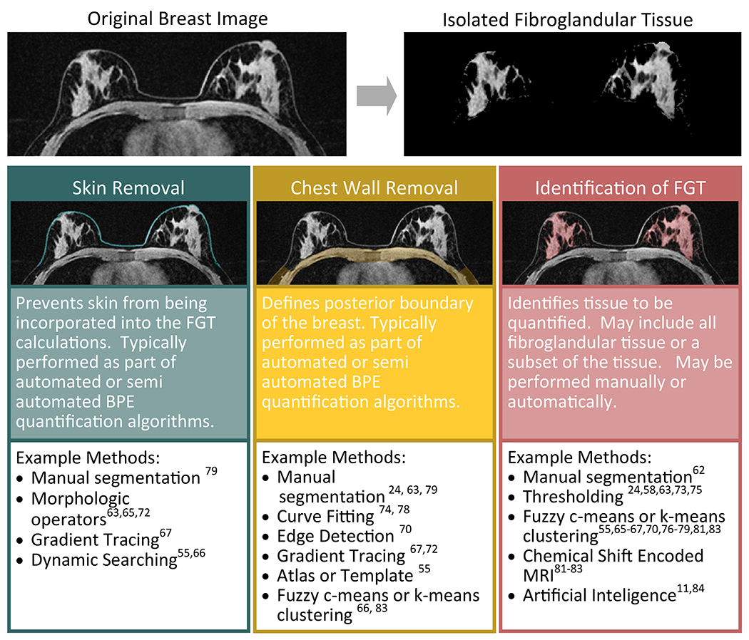
Diagram of the potential steps involved in isolation of the fibroglandular tissue (FGT) for quantification of BPE. Many different approaches have been taken to isolate the FGT ranging from pure manual segmentation to fully automatic algorithms. Typical steps, illustrated here, include skin removal, chest wall removal, and final isolation of the FGT itself. These steps may be performed on a single representative slice of the breast or on the entire breast volume.