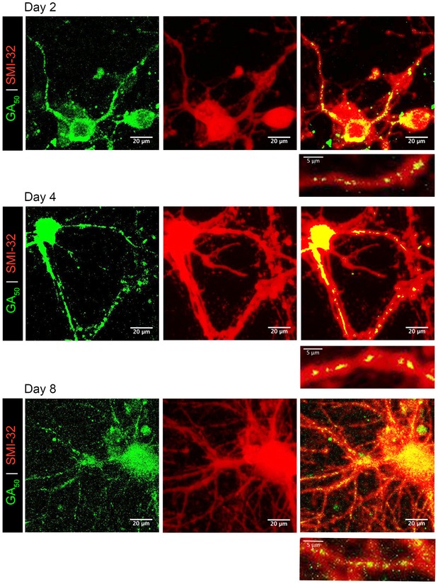
Figure 1. GA 50 aggregates are detected in neurites of cortical neurons over time.

Primary rat cortical neurons transfected with eGFP‐GA50 were examined to determine at which time points preceding cell‐death aggregates are found in neurites. Two days post‐transfection, aggregates formed by eGFP‐GA50 (green) are detectable in neurites (SMI‐32 staining in red). These aggregates remain localized to neurites at 96 h (4 days) and 288 h (8 days). Colocalization is indicated by yellow overlay of colors (right panels). Inset below each image shows enlargement of neurite regions containing aggregates, representative fields from 60× magnification z‐stack confocal images, scale bar indicates 20 μm. Inset scale bars indicate 5 μm.
