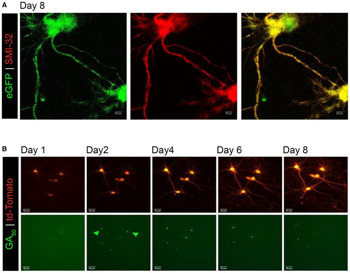
Figure EV1. GA aggregates are dynamic.

-
AeGFP was expressed in mature cortical neurons for 8 days and then immunostained for neurofilament (SMI‐32, red) and eGFP (green). This representative z‐stack confocal images demonstrate cellular viability and lack of GFP aggregation when expressed in the absence of GAn dipeptides even at this extended time. 60× magnification, scale bar indicates 20 μm.
-
BPrimary rat neurons were co‐transfected with Td‐tomato and eGFP‐GA50 plasmid. The same neurons were imaged at 24‐h intervals. Representative fields of td‐Tomato (top) and eGFP‐GA50 (bottom) co‐positive cortical neurons at Days 1, 2, 4, 6, and 8 post‐transfection follow individual cells over time. Highlighted with green arrows are GA aggregates that dissipate over the course of our imaging period, while the cells containing them remain viable. 20× magnification, scale bar indicates 40 μm.
