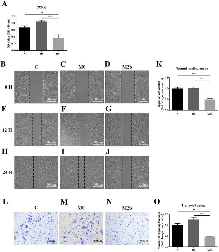
Figure 2. Conditioned medium from M2b macrophages inhibited the proliferation and migration of PASMCs.
PASMCs were treated with serum-free medium, supernatant of M0 macrophages, and supernatant of M2b macrophages for 24 hours, and the proliferation and migration of the different groups were detected. (A) Proliferation of PASMCs detected by CCK-8 assay (n = 6 for each group). (B–K) Migration of PASMCs detected by wound healing assay (n = 5 for each group, original magnification: 50×). (L–O) Migration of PASMCs detected by transwell assay (n = 5 for each group, original magnification: 100×). Data are shown as the mean ± SEM. **p < 0.01, ***p < 0.001. “C” indicates the control group, “M0” indicates the M0 macrophage group, “M2b” indicates the M2b macrophage group.

