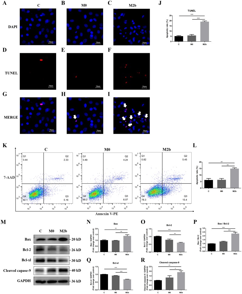
Figure 3. Conditioned medium from M2b macrophages attenuated the apoptosis resistance of PASMCs.
PASMCs were treated with serum-free medium, the supernatant of M0 macrophages or the supernatant of M2b macrophages for 24 hours before detection. (A–J) TUNEL staining was used to assess the apoptosis of PASMCs (n = 5 for each group, original magnification: 200×). The total number of PASMCs counted across the n = 5 was as follows: C group = 207, M0 group = 177, M2b group = 188. The white arrow points to rippled or creased nuclei. The apoptotic cell proportion was calculated as the ratio of TUNEL-positive cells to the total number of PASMCs. (K, L) Annexin V-PE/7-ADD staining was used to assess the apoptosis of PASMCs (n = 3 for each group). The number of apoptotic cells was quantified by flow cytometry after the cells were stained with annexin V-PE and 7-ADD. Early apoptotic cells were determined by counting the percentage of annexin V (+), 7-ADD (−). (M–R) Western blot was then used to assess the protein expression of Bax, Bcl-2, Bcl-xl, cleaved caspase-9, and GAPDH (n = 4 for each group). Data are shown as the mean ± SEM. *p < 0.05, **p < 0.01, ***p < 0.001. “C” indicates the control group, “M0” indicates the M0 macrophage group, “M2b” indicates the M2b macrophage group.

