
Figure 2 |.
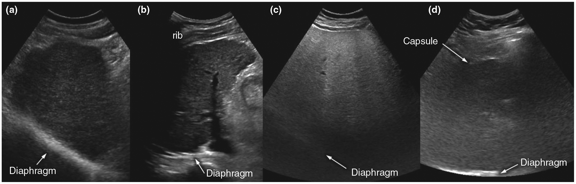
Illustrative examples of adequate and inadequate ultrasound quality. (a) Adequate-quality exam: Although diffusely heterogeneous, liver parenchyma is clearly visualised and focal liver lesions were ruled out with high confidence. (b) Inadequate-quality exam: Right hepatic dome could not be visualised due to extensive rib shadowing. (c) Inadequate-quality exam: Posterior half of the liver could not be visualised due to severe parenchymal fatty liver disease. (d) Inadequate-quality exam: Liver parenchyma is poorly visualised throughout due to morbid obesity and thick subcutaneous/visceral fat, in addition to probable severe underlying parenchymal disease.