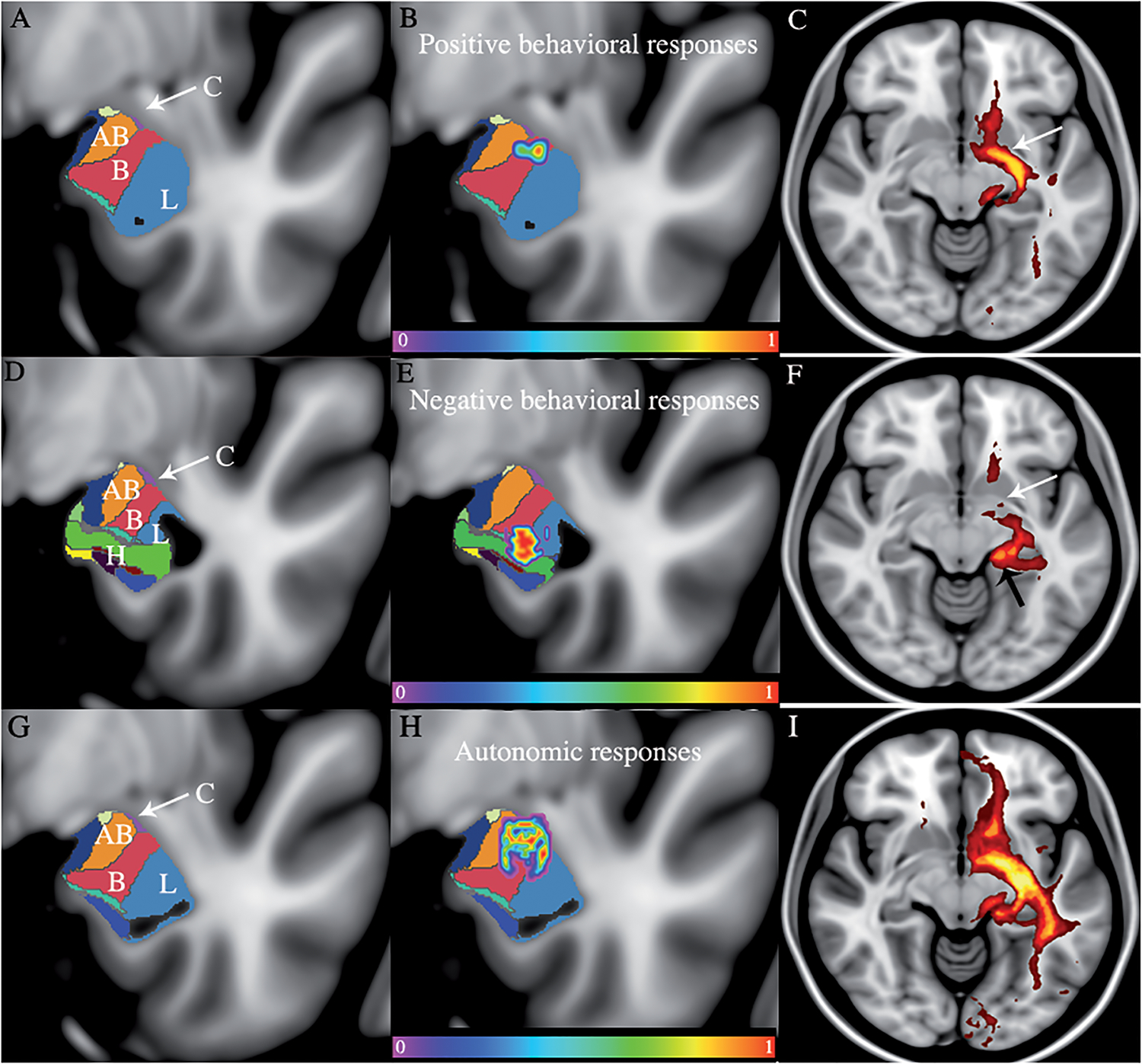
Figure 1: Structural mapping of the behavioral responses after DBS of the basolateral amygdala.

The averaged clusters of both subjects were overlaid with the histological segmentation of the amygdala. A-D-G. The entire stimulated area included the accessory basal nucleus (AB), the basal nucleus (B), the lateral nucleus (L), the central nucleus (C with arrow), and the hippocampus (H). B. The averaged cluster associated with the positive behavioral responses is located in the basal nucleus. C. The positive behavioral responses were associated with involvement of the ventral amygdalofugal pathway (VAF) E. The averaged cluster associated with negative behavioral responses is mainly located in the lateral nucleus and the hippocampal area. F. The negative responses mainly involved the stria terminalis (ST) H. The averaged cluster associated with autonomic responses is located from the most dorsal to the central part of the amygdala involving the white matter dorsal to the amygdala, the basal, accessory basal, lateral, and the central nucleus of the amygdala. I. The autonomic responses involved the ST and the VAF. The MNI coordinates of the coronal slices are as follows: Positive behavioral responses B: y=−5. Negative behavioral responses E: y=−8. Autonomic behavioral responses H: y=−6.
