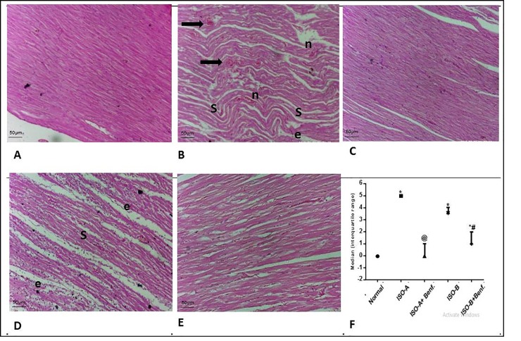
Fig 5. Effect of benfotiamine pre- and post-treatments on histological damage in ISO-induced MI in rats.
(A–F) Cardiac specimens were stained with H&E (magnification x200). Normal group (A) showed normal histological structure of cardiac muscle. ISO-A treatment group (B) showed severe cardiac insult with atrophy, muscle shrinkage (s) edema (e) of cardiac muscle, marked inflammatory cells infiltration (n), extensive edema in-between muscle fibers (e), and marked number of apoptotic or degenerated myocytes filament (black arrows). Benfotiamine pre-treatment group (C) showed mostly normal cardiac muscle architecture. ISO-B treatment group (D) revealed shrinkage (s) of cardiac muscle with preserved nuclei all over sections examined, edema in between muscle fibers (e). Benfotiamine post-treatment (E) demonstrated almost normal cardiac muscle with only mild edema in-between muscle fibers. Myocardial score of damage expressed as median (interquartile changes) (F). Each value represents the median value [interquartile range] (n = 6). Statistical analysis was done using non-parametric One-Way ANOVA (Kruskal-Wallis test) followed by Dunn's multiple comparison test. * p < 0.05 vs. normal, @ p < 0.05 vs. ISO-A, # p < 0.05 vs. ISO-B. ISO-A: ISO pre-treatment control; ISO-B: ISO post-treatment control; ISO-A+ Benf.: benfotiamine prophylactic; ISO-A+ Benf.: benfotiamine treatment.

