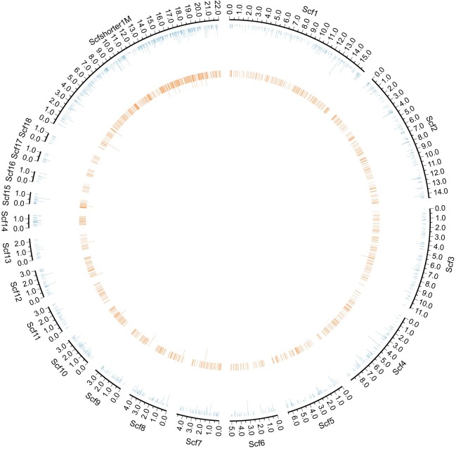
Fig 3. The distribution of cell-free E. granulosus DNA reads on the nuclear genome.
The circulation genome visualization showed the E. granulosus reads mapping position on the nuclear genome (outermost blue circle). Eighteen scaffolds longer than 1Mb were displayed in the separate fragments (Scf1-Scf18). Scaffolds shorter than 1Mb were concatenated to display (Scfshort1M). The inner orange circle represents the count of patients with reads detected in the region. Circle figures of the E. granulosus mitochondrial genome were put in the supplementary materials (S1 Fig).

