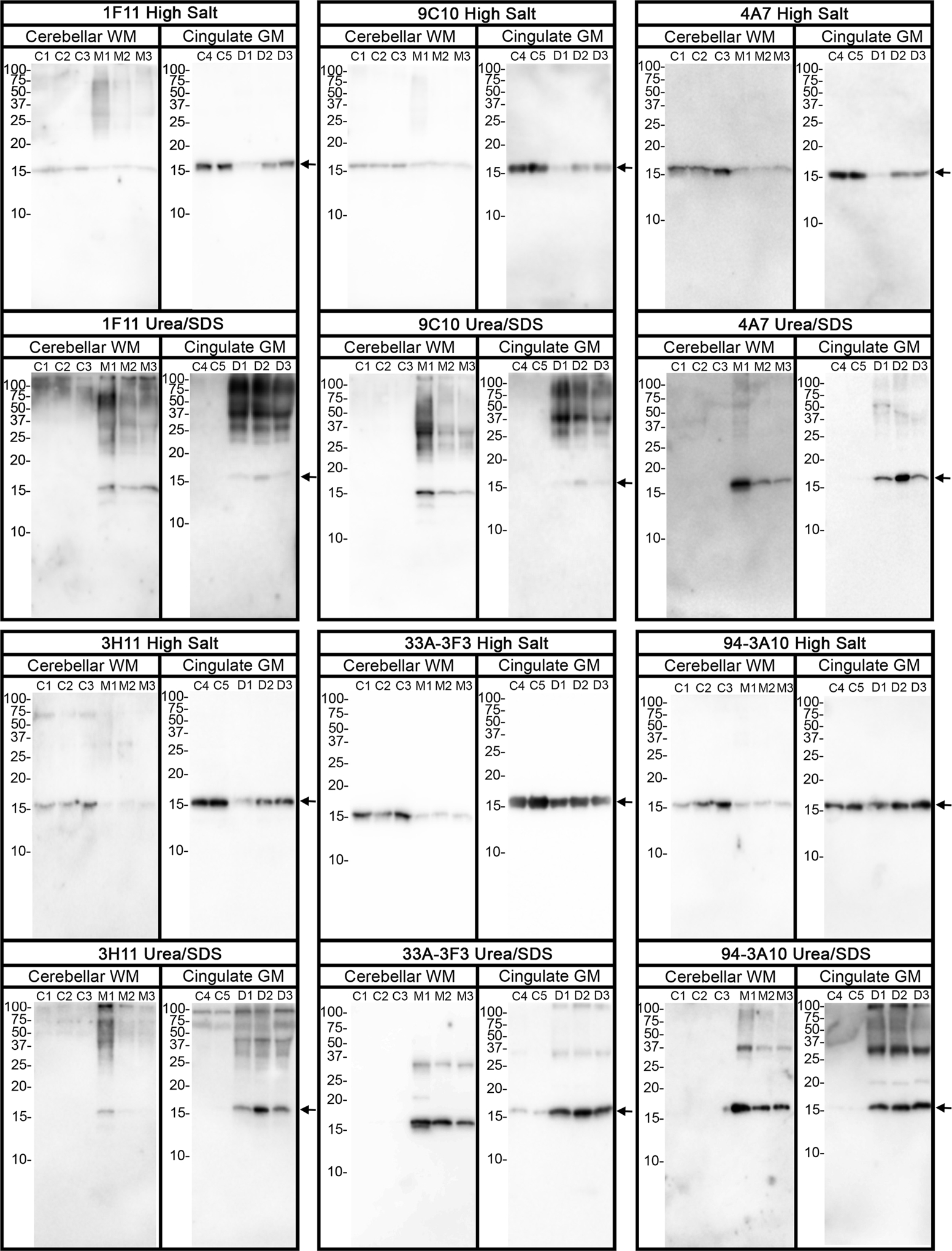
Figure 3. Biochemical comparisons of αS immunological profiles in MSA and DLB brain specimens.

Immunoblot comparison of αS found in the cerebellum white matter of MSA case 2 (lane M1), case 3 (lane M2), and case 4 (lane M3), with αS found in cingulate cortex of three confirmed DLB patients (D1, D2, and D3). Control samples C1, C2, and C3 come from cerebellar white matter (WM) of patients with no neurodegenerative phenotypes, and control sample C4 and C5 come from cingulate cortex gray matter (GM) of patients with no neurodegenerative phenotypes. Samples were sequentially fractionated as described in “Materials and Methods”. Soluble high salt compared to detergent insoluble and urea/SDS samples from each case were loaded on separate lanes of SDS-polyacrylamide gels that were then analyzed by immunoblotting with the respective αS antibodies as indicated above each panel. The mobilities of molecular mass markers are indicated on the left of each blot. The arrows on the right depict monomeric αS.
