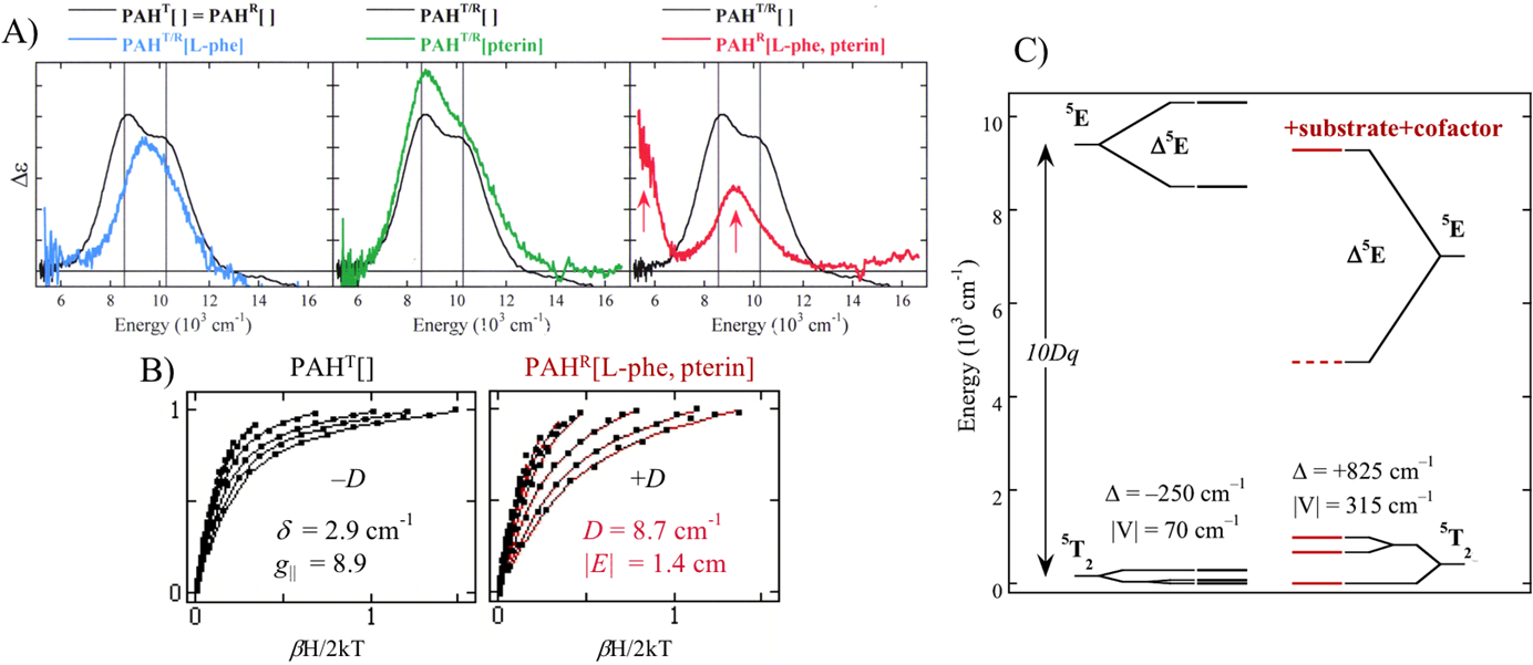
Fig. 4.

A) LT MCD spectra of resting PAH (black), substrate-bound PAH (blue), cofactor-bound PAH (green) and substrate plus cofactor bound PAH (red). B) Saturation magnetization data for resting PAH (black, left) and substrate plus cofactor bound PAH (red, right). C) VTVH MCD obtained LF splitting of d-orbitals in resting PAH (black, left) and substrate plus cofactor bound PAH (red, right). Adapted from Ref. 9.
