Figure 5.

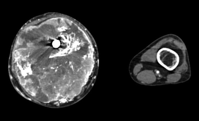
Photograph showing CT contrast-enhanced transaxial imaging demonstrating a large soft-tissue lesion with necrosis and osseous destruction of the distal femur.

Photograph showing CT contrast-enhanced transaxial imaging demonstrating a large soft-tissue lesion with necrosis and osseous destruction of the distal femur.