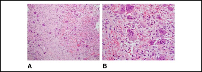
Figure 7.
A, Micrograph showing a tumor composed of a haphazard and fascicular proliferation of bland spindle cells and osteoclast-type giant cells. Giant cells have an uneven distribution and cluster around the areas of hemorrhage (100×, H&E). B, Micrograph showing spindle cells with a disorderly arrangement with extravasated red blood cells and giant cells. The nuclear features of the spindle cells and giant cells are distinct from one another and significant atypia is absent (400×, H&E).

