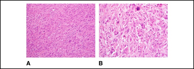
Figure 8.
A, Photograph showing microscopically markedly atypical spindle cells arranged in fascicles with brisk mitotic activity including atypical forms. Scattered tumor giant cells and epithelioid cells are also present (200×, H&E). B, Photograph showing other areas show more sheet-like growth with elongated and polygonal cells. Tumor giant cells and atypical mitotic figures are conspicuous (400×, H&E).

