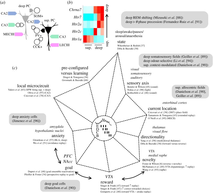
Figure 3.
Biases of hippocampal replay can affect deep and superficial (sup.) CA1 pyramidal cells differently. (a) Known deep superficial local microcircuit motifs. Deep cells are more strongly activated by inputs from CA2 (at basal dendrites) and the medial entorhinal cortex. Inputs from CA3 cells onto deep cells are strongly interfaced by feed-forward inhibition. Superficial cells receive more innervation from the lateral entorhinal cortex and direct CA3 inputs. Importantly, lateral and medial entorhinal inputs to deep and superficial cells organize proximodistally along the traverse CA1 axis. Superficial cells mainly recruit PV+ basket cells whereas deep cells are biased for SOM+ interneurons. In return, innervation by PV+ basket cells is larger over deep cells while CCK+ basket cells preferentially target superficial cells. (b) Differential transcriptomic expression of serotoninergic (Htr) and cholinergic receptor genes (Chrn) along the deep and superficial layers. Normalized gene expression values from three different replicates are shown. Data from https://hipposeq.janelia.org/ [92]. (c) Multifactorial axes biasing the content and organization of replays during sharp-wave ripples. Different influences on deep and superficial CA1 pyramidal cells have been reported along these axes (grey boxes). The relative axis length is not necessarily informative [6,30,34,36–38,40,43,45–51,57,58,68,78,88–91,93–98]. PC, pyramidal cell, SOM+, somatostatin-positive interneuron; PV+, parvalbumin-positive basket cell; CCK+, cholecystokinin-positive basket cell; MECIII, medial entorhinal cortex layer III; LECIII, lateral entorhinal cortex layer III. (Online version in colour.)

