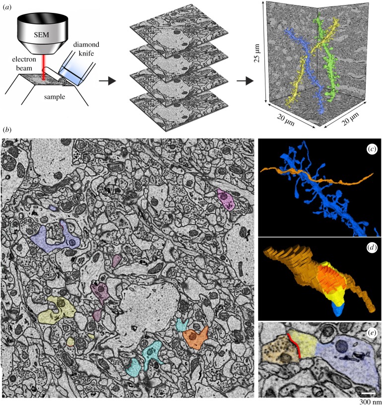
Figure 2.
(a) Schematic showing the basic principle of serial block-face scanning electron microcopy: the face of the sample block is imaged and sliced repeatedly. Consecutive images are collected in stacks that are automatically aligned and then used for the manual segmentation of the structures of interest; for instance, the three dendritic branches shown in colour on the right (CA1 stratum radiatum, mouse P30; [32]). SEM, scanning electron microscope. (b) Example of one two-dimensional image in the stack where several dendritic elements and spines were manually segmented (CA1 stratum radiatum, mouse P30; [32]. Each dendrite and its spines are indicated by a different colour. Trained annotators learned to use the open source software Fiji to manually trace the object of interest (spine, ASI, axon bouton, etc.) over serial sections, to obtain a three-dimensional reconstruction of the object. All final reconstructions were checked for accuracy and consistency by the same person, to ensure that all annotators followed the same established rules. (c,d) Three-dimensional representation of a dendritic segment (in blue) and the axon making synaptic contact (orange); the lower panel shows one spine head (yellow) contacted by the axon (orange); their direct area of contact, the ASI, is indicated in red. (e) Two-dimensional image showing an example of a segmented axon bouton with synaptic vesicles (orange), ASI (red), spine head (yellow) and dendritic segment (blue).

