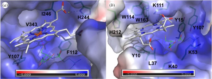
Figure 4.
Comparison of the active sites from mPGES2 bound to haem and AtGSTU23 docked with protoporphyrin IX. (a) Crystal structure of human mPGES2 bound to GSH and haem (PDB code 2PBJ). (b) Docking of PPIX onto AtGSTU23 crystal structure (PDB code 6EP7). Molecular surfaces coloured according to the electrostatic potential were calculated with APBS software [70] and are shown in transparency. PPIX and haem are represented as white sticks. Residues that line the active sites are shown as green sticks. Putative H-bonds are shown as yellow dashed lines. Coordination bond between haem and GSH is shown as pink dashed line. GSH is shown as green lines.

