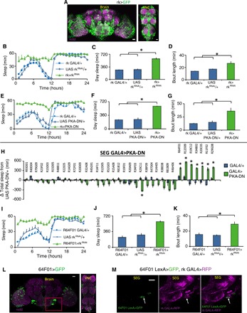
Fig. 3. rickets neurons regulate wing expansion and sleep.

(A) rk-GAL4/+>UAS-GFP/+ labels a large number of cells in the fly CNS. (B and E) Sleep was increased in both rk-GAL4/+>UAS-rkRNAi/+ and rk-GAL4/+>UAS-PKADN/+ flies compared to parental controls (n = 16 to 32 flies per genotype; repeated-measures ANOVA for Time × Genotype, P < 0.001). rk-GAL4/+>UAS-rkRNAi/+ and rk-GAL4/+>UAS-PKADN/+ displayed increased daytime sleep (C and F) and sleep bout duration (D and G) compared to controls (*P < 0.01, Tukey correction). (H) Screen for SEG GAL4 drivers that increase sleep when expressing UAS-PKADN; sleep is expressed as change in sleep in minutes relative to the UAS-PKADN/+ controls (*P < 0.01, Tukey correction). The names of the GAL4 lines tested are listed above. (I) 64F01-GAL4/+>UAS-rkRNAi flies with unexpanded wings slept more than parental controls (n = 16 to 32 flies per genotype; repeated-measures ANOVA for Time × Genotype, P < 0.001). (J and K) 64F01-GAL4/+>UAS-rkRNAi flies displayed increased daytime sleep and sleep bout duration compared to controls (*P < 0.01, Tukey correction). (L) R64F01-GAL4/+>UAS-GFP/+ labels a sparse population of cells in the CNS, including the SEG (red box). (M) R64F01LexA/+>LexAopGFP/+; rk-GAL4/+>UAS-RFP/+ (red fluorescent protein) overlap in one cell in the SEG (white arrow). (L) Maximal intensity z projections counterstained with nc82 (magenta). (M) Single confocal slices. Scale bar, 20 μm.
