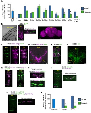
Fig. 5. Circuit for wing cut–induced sleep.

(A) Change in sleep (cut-intact) in flies expressing UAS-Kir2.1 in GAL4 lines associated with wing chemo- and mechanosensation normalized to the UAS-Kir2.1 parental control. Ir52a-GAL4/+>UAS-Kir2.1/+ and Ir76b-GAL4/+>UAS-Kir2.1/+ flies displayed reduced sleep in response to wing cut relative to controls (n = 20 to 45 flies per condition; *P < 0.05, Tukey correction). Ir76GAL4/+; Ir52a-GAL4/+>UAS-Kir2.1/+ flies did not increase sleep in response to wing cut. (B) Ir52a-GAL4/+>UAS-GFP/+ labels subsets of wing neurons (left) that project into the wing neuromere of the VNC (middle). Weak expression was also detected in nerves from leg neurons that project into the VNC (middle). No expression was detected in the brain (right). (C) Ir52a-GAL4/+>Trans-tango/+ (magenta) detects neurites in close proximity to the projections of Ir52a-GAL4 axons (green) in the VNC, with prominent labeling in the wing neuromere (white arrow), and two projection neuron axon tracts that exit the VNC and project to the lateral protocerebrum. (D) Ir52a-GAL4/+>Trans-tango/+ labels VNC neurons that project axons out of the VNC into the brain in two tracts with arborizations in the SEG and the VLP (orange and yellow arrows). (E) In the VNC, 31C06-GAL4/+>UAS-GFP labels neurites that resemble the Ir52a>trans-tango pattern in (C) with strong labeling in the wing neuromere (white arrow). (F) In the brain, 31C06-GAL4/+>UAS-GFP/+ labels neurons that project in patterns similar to the Ir52a>trans tango–labeled axons (orange and yellow arrows, “1” and “2”). (G and H) 31C06-GAL4/+>UAS-Denmark,UAS syt_EGFP/+ and Ir52a-GAL4/+>UAS-Denmark,UAS syt:EGFP/+ staining patterns. UAS-Denmark (magenta) labels dendrites; syt:EGFP (green) labels presynaptic sites. (I) 31C06LexA/+>LexAop CD8:GFP/+ and Ir52a-GAL4/+>UAS CD8:RFP/+ expression patterns reveal that 31C06 dendrites (GFP, green) are in close proximity to Ir52a-GAL4 axons (RFP, red), particularly in the wing neuromere (right). (J) Strong GRASP signal was detected between 31C06LexA dendrites and Ir52a-GAL4 axons in the VNC (left). GRASP signal was in close proximity to Ir52a-GAL4 presynaptic sites (right, brp:mcherry in magenta). (K) 31C06-GAL4/+>UAS-Kir2.1 and 17F09GAL4/+>UAS-Kir2.1 blocked the increase in sleep following wing cut compared to parental controls (n = 27 to 46 flies per condition; *P < 0.01, Tukey correction). (B to J) Maximum intensity confocal projections. Wing neuromere image to the right in (J) is a single confocal slice. Scale bar, 20 μm.
