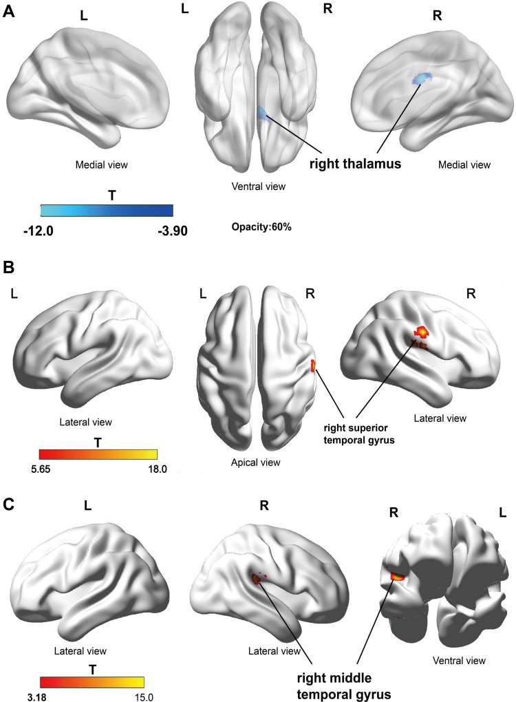
Figure 4.
DKI analyses suggest brain structural changes in PHN patients after treatment. (A) AD value of right thalamus (Thalamus_R (aal)) was significantly reduced after treatment; (B) FA values in the right superior temporal gyrus (Temporal_Sup_R (aal)) increased after treatment; (C) AK values in the right temporal gyrus (Temporal_Mid_R (aal)) increased after treatment (P <0.05, AlphaSim correction). The blue color indicates the GMV is significantly reduced after treatment, and the red and yellow colors indicate the GMV is significantly increased after treatment.
Abbreviation: aal, anatomical automatic labeling.

