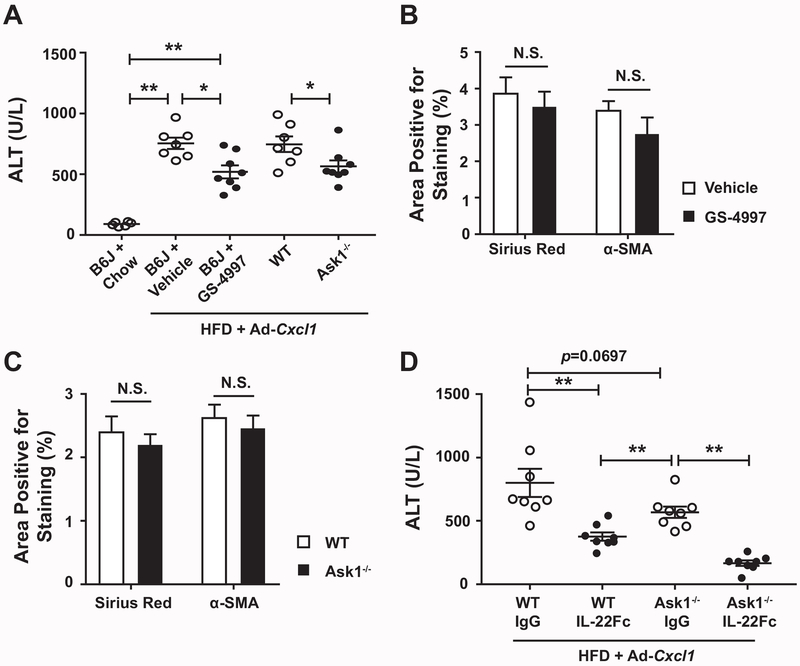
Fig. 4. ASK1 inhibition fails to ameliorate CXCL1-induced NASH and IL-22Fc improves NASH in an ASK1-independent manner.
(A-C) HFD-fed C57BL/6J mice (B6J), Ask1−/− mice and their WT littermates were infected with Ad-Cxcl1 for two weeks. B6J group was also given vehicle or GS-4997 treatment (n=7–8/group). (A) Serum ALT levels. (B) Quantification of the histological analysis of Sirius Red and α-SMA in liver sections. The representative images are included in Supporting Fig. S11. (C) Quantification of the histological analysis of liver fibrosis in Ask1−/− mice and WT littermates. The representative images are included in Supporting Fig. S12. (D) Ask1−/− mice and WT littermates were given HFD + Ad-Cxcl1, followed by a treatment with IgG2 or IL-22Fc (n=8/group). Serum ALT levels were measured. Values represent mean ± SEM. Statistical evaluation was performed by Student’s t-test or one-way ANOVA with Tukey’s post hoc test for multiple comparisons (*p<0.05; **p<0.01).

