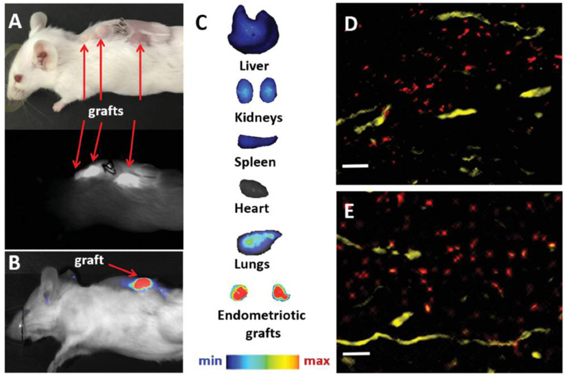
Figure 6.
A) Photograph (top) and NIR fluorescence image (bottom), recorded with Fluobeam 800, of a mouse bearing endometriotic grafts 24 h after intravenous injection of “activatable” SiNc-NP. B,C) NIR fluorescence images of a mouse bearing endometriotic graft (B) and resected tissues (C) recorded with Pearl Impulse Small Animal Imaging System 24 h after intravenous injection of “activatable” SiNc-NP. D,E) Representative fluorescence microscopy images of sections of endometriotic grafts collected 24 h after intravenous injection of SiNc-NP. Red color indicates NIR fluorescence generated by SiNc-NP. Yellow color represents blood vessels stained with the fluorescently labeled anti-CD31 antibody. Scale bars are 50 μm.

