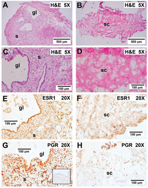
Figure 8.
Effect of PTT on graft histology and steroid receptor staining. A,C) In control mice not receiving PTT, the grafts displayed enlarged endometriotic glands and stroma. E,G) In these mice, the grafts stained strongly for both ESR1 and PGR. B,D) Endometriotic glands and stroma were lost after PTT and graft sites were replaced with murine connective tissue. F,H) Minimal ESR1 and PGR staining nuclei were observed after PTT therapy. The inset in G shows irrelevant IgG control (anti-Br(d) U) showing staining specificity for both ESR1 and PGR. “gl” indicates glands; “s” indicates stroma; “sc” indicates stromal connective tissue with minimal ESR1 and PGR staining. 5X, 5× plan apochromatic objective; 20X, 20× plan apchromatic objective.

