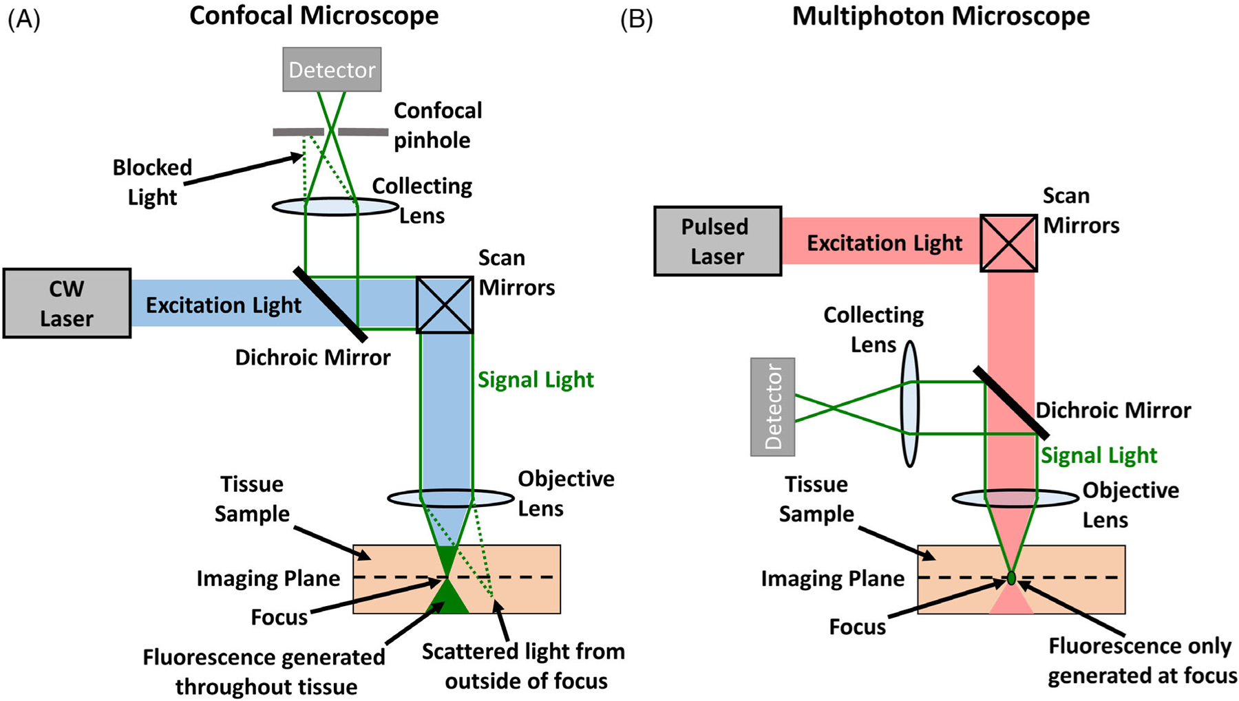
Figure 2.

Confocal and multiphoton microscopes capture images from a single plane within solid tissue. A, Optical layout of a confocal microscope. Though the continuous wave (CW) laser generates signal throughout the tissue sample, out of focus light is eliminated by focusing returning signal light through a pinhole with a collecting lens. Light originating from within the focal point of the objective lens passes through the pinhole and can reach the detector. Light originating outside of the focus is blocked by the pinhole. B, Optical layout of a multiphoton microscope. The use of a femtosecond pulsed laser allows multiphoton excitation of the sample and signal light is only generated at the exact focus. In this case, a pinhole is not required and all signal can be collected on the detector. In both confocal and multiphoton microscopy, signal from the entire imaging plane is generated by scanning the focal point over the imaging plane with the scan mirrors.
