
Figure 2. Upregulated PD-L1 by HDAC inhibition was due to the increased CD274 transcription.
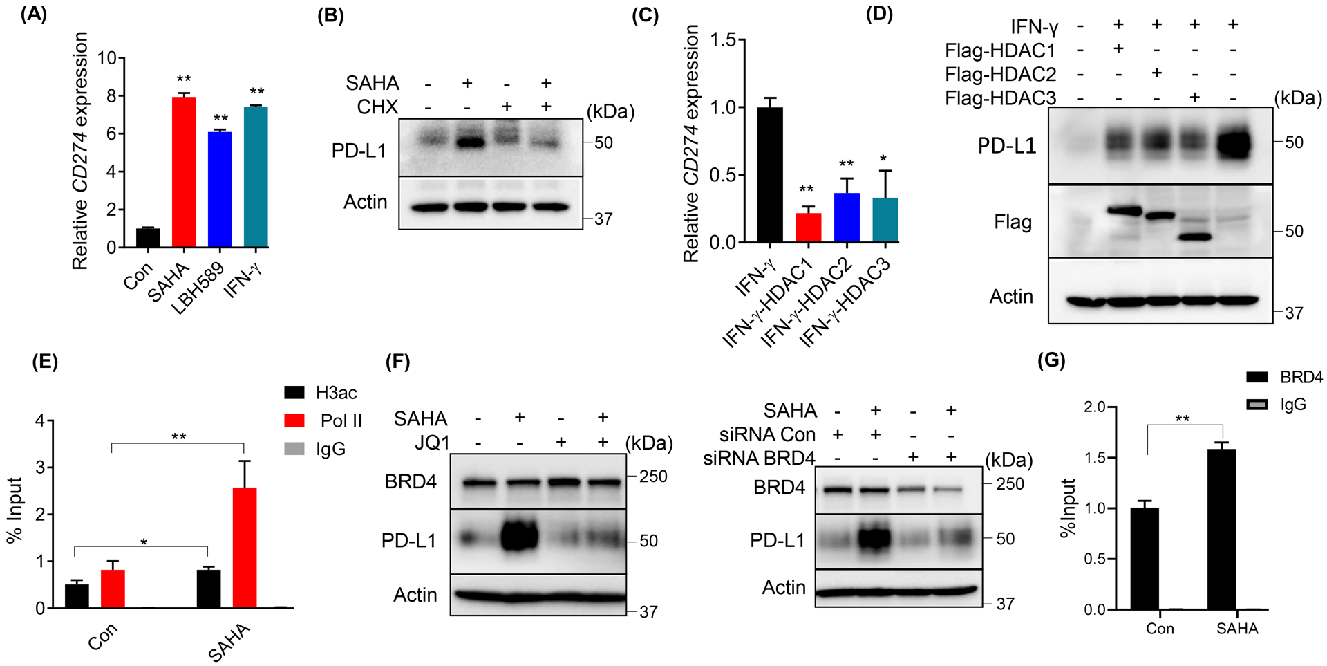
(A) Expression of CD274 was analyzed by RT-qPCR after DU145 cells were treated with 1 μM SAHA or 50 nM LBH589 or 10 ng/ml IFN-γ for 24 h. (B) IB analysis of WCL derived from DU145 cells as indicated treated (100 μg/ml CHX or 1 μM SAHA) for 12 h. (C, D) DU145 cells were transfected with the indicated constructs, followed with 10 ng/ml IFN-γ, and harvested to measure mRNA levels (C) or protein levels of PD-L1 (D). (E, G) ChIP-qPCR analysis of H3ac, BRD4 and Pol II binding at CD274 promotor in DU145 cells treated with 1 μM SAHA. (F) IB analysis of WCL derived from DU145 cells treated with 1 μM SAHA and 5 μM JQ1 (left panel) or transfected with siRNA to deplete BRD4, then treated with 1 μM SAHA (right panel). The statistical data were shown as mean values ± SD (n ≥ 3), *p < 0.05 **p<0.01.